mail_outline sales@mediastorehouse.com
Mole - eating worm undergoundJD-13175 European / Common MOLE - eats worm in hole underground Talpa europaea John Daniels Please note that prints are for personal display purposes only and may not be reproduced in any way
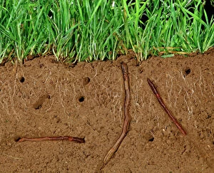
Earthworms - Soil cross-section showing worms in tunnnels and aeration and grass roots JPF12192JPF-6743 Earthworms - Soil cross-section showing worms in tunnnels and aeration and grass roots Jean Paul Ferrero Please note that prints are for personal display purposes only
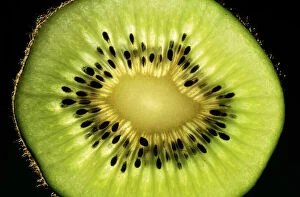
Kiwi Fruit - cross section showing seedsDOW-23 KIWI Fruit - cross section showing seeds Steve Downer Please note that prints are for personal display purposes only and may not be reproduced in any way
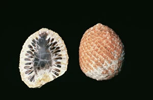
Fossil Araucaria Cones, cross section & full cone, Mid Jurassic, Province Santa Cruz, ArgentinaFG-11972 Fossil - Araucaria Cones, cross section & full cone Mid Jurassic, Province Santa Cruz, Argentina. Francois Gohier Please note that prints are for personal display purposes only
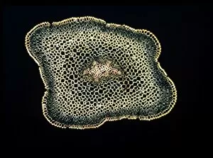
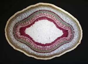
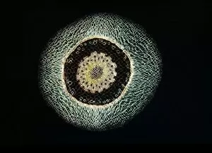
Light Micrograph (LM): A transverse section of a stem of Whisk Fern (Psilotum nudum); Magnification x18 (on 10)LRDS-167 Light Micrograph (LM): A transverse section of a stem of Whisk Fern (Psilotum nudum) Magnification x18 (on 10.5 cm width print) Psilotum nudum David Spears (Last Refuge)
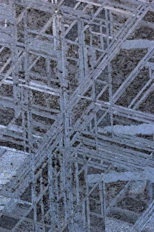
Meteorite Cross-section - Muonionalusta-Kirunu Norbotten Sweden -Found in 1906- Iron-Octahedrite-fineCAN-2438 Meteorite Cross-section Muonionalusta - Kirunu Norbotten Sweden - Found in 1906 - Iron - Octahedrite - fine John Cancalosi Please note that prints are for personal display purposes only
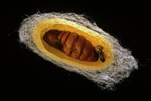
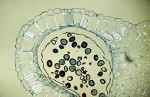
Silk Moth - cross section of cocoonPPG-180 Silk Moth - cross section of cocoon Bombyx mori Pascal Goetgheluck contact details: prints@ardea.com tel: +44 (0) 20 8318 1401
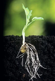
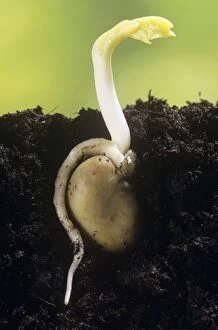
Broad Bean - showing root growthJD-16083 Broad BEAN - shows roots growing John Daniels Please note that prints are for personal display purposes only and may not be reproduced in anyway
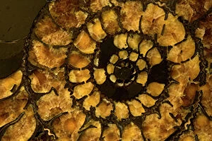
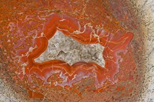
Fossil Ammonite Cross section (Speetoniceras) - Russia - CretaceousCAN-2455 Fossil Ammonite Cross section (Speetoniceras) Russia - Cretaceous John Cancalosi Please note that prints are for personal display purposes only and may not be reproduced in any way
Broad Bean - showing rootJD-15828 Broad Bean - shoot appearing above soil Underground, c/u John Daniels Please note that prints are for personal display purposes only and may not be reproduced in anyway
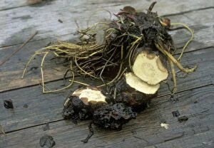
Water Lily crown rot shows as brown tissue in the rhizomesBB-1341 Water Lily crown rot shows as brown tissue in the rhizomes Brian Bevan Please note that prints are for personal display purposes only and may not be reproduced in anyway
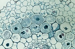
Light Micrograph (LM): Transverse section of Dehiscence Lilium Anthers with Pollen; Magnification x300 (on 10)LRDS-200 Light Micrograph (LM): Transverse section of Dehiscence Lilium Anthers with Pollen Magnification x300 (on 10.5 cm width print) David Spears (Last Refuge)
Mammoth Tusk - Cross section - North Yakutia - Russia - 25, 000 years oldCAN-2488 Mammoth Tusk - Cross section North Yakutia - Russia - 25, 000 years old John Cancalosi Please note that prints are for personal display purposes only and may not be reproduced in any way
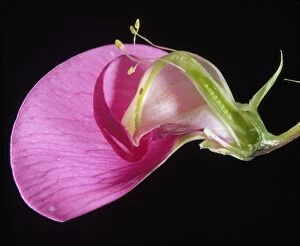
Sweet Pea - sectionLA-105 Sweet Pea - section Jean Michel Labat Please note that prints are for personal display purposes only and may not be reproduced in anyway
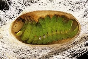
Indian Moon Moth Caterpillar - Cross section of caterpillar in cocoon prior to pupationPPG-234 Indian Moon Moth Caterpillar - Cross section of caterpillar in cocoon prior to pupation Actias selene Pascal Goetgheluck contact details: prints@ardea.com tel: +44 (0) 20 8318 1401
Agatized Dinosaur Bone - Allosaur - Cross section shows bone marrow of femur - Amethyst crystal centers - Late JurassicCAN-3448 Agatized Dinosaur Bone - Allosaur Cross section shows bone marrow of femur - Amethyst crystal centers - Late Jurassic - Utah John Cancalosi contact details: prints@ardea.com tel
Light Micrograph (LM): Filaments of fungi Endotrophic Mycorrhiza live within cells of a root; Magnification x600LRDS-254 Light Micrograph (LM): Filaments of fungi Endotrophic Mycorrhiza live within cells of a root Magnification x600 (on 10.5 cm width print) David Spears (Last Refuge)
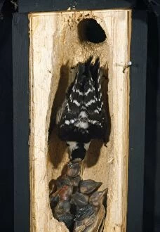
Lesser-spotted Woodpecker - cross section of tree into nest with adult feeding youngWOM-44 Lesser-spotted Woodpecker - cross section of tree into nest with adult feeding young Dendrocopos minor Wilhelm Moller Please note that prints are for personal display purposes only
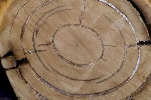
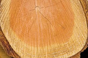
Douglas Tree - Growth ring The Netherlands, Overijssel, forestry StaphorstVG-1601 Douglas Fir Tree - Growth ring The Netherlands, Overijssel, forestry Staphorst Pseudotsuga menziesii Paul Van Gaalen Please note that prints are for personal display purposes only
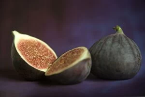
Fig - in studioLA-4256 Fig - in studio Jean Michel Labat Please note that prints are for personal display purposes only and may not be reproduced in any way
Earthworm pulling leaf down into its burrow KAT01964AUS-1938 Earthworm - pulling leaf down into its burrow Kathie Atkinson / Auscape / ardea.com Auscape Please note that prints are for personal display purposes only
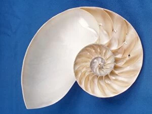
Nautilus - sectionPJG-339-c Nautilus - section Pompilius philippians Peter Green Please note that prints are for personal display purposes only and may not be reproduced in any way
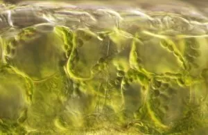
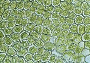
Light Micrograph (LM): plant cell chloroplasts - the site where photosynthesis takes placeLRDS-192 Light Micrograph (LM): plant cell chloroplasts - the site where photosynthesis takes place David Spears (Last Refuge)
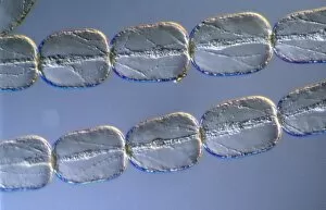
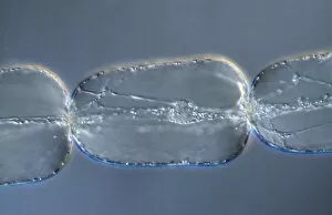
Light Micrograph (LM): Cells from a hair on the stamen of the common spiderwort (Tradescantia)LRDS-171 Light Micrograph (LM): Cells from a hair on the stamen of the common spiderwort (Tradescantia) Tradescantia sp David Spears (Last Refuge)
Light Micrograph (LM): A transverse section of a Fig leaf); Magnification x15 (on 10. 5 cm width print)LRDS-187 Light Micrograph (LM): A transverse section of a Fig leaf) Magnification x15 (on 10.5 cm width print) David Spears (Last Refuge)
Light Micrograph (LM): The cellular struture of a liverwort plant (Hepatica); Magnification x1200 (on 10)LRDS-190 Light Micrograph (LM): The cellular struture of a liverwort plant (Hepatica) Magnification x1200 (on 10.5 cm width print) Hepatica sp David Spears (Last Refuge)
Light Micrograph (LM): A single cell from a hair on the stamen of the common spiderwort (Tradescantia)LRDS-170 Light Micrograph (LM): A single cell from a hair on the stamen of the common spiderwort (Tradescantia) Tradescantia sp David Spears (Last Refuge)
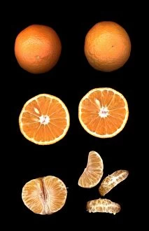
Fruit - MandarinWAT-12573 Fruit - Mandarin M. Watson Please note that prints are for personal display purposes only and may not be reproduced in anyway. contact details: prints@ardea.com tel: + 44 (0) 20 8318 1401
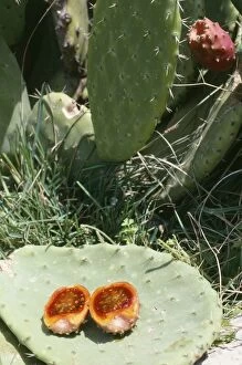
Prickly Pear Cactus - showing fruit in sectionSG-7708 Prickly Pear Cactus - showing fruit in section Ardea London Please note that prints are for personal display purposes only and may not be reproduced in anyway
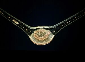
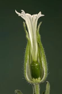
White Campion - cross-section of a female flower EuropePPG-1490 White Campion - cross-section of a female flower Europe Silene alba Pascal Goetgheluck Please note that prints are for personal display purposes only and may not be reproduced in anyway
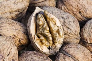
Walnuts - closeup of basket with single fruit opened to expose the edible walnut seedMAB-440 Walnuts - closeup of basket with single fruit opened to expose the edible walnut seed. Juglans regia Mark Boulton Please note that prints are for personal display purposes only
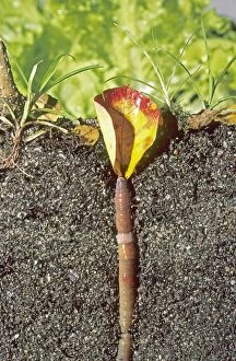
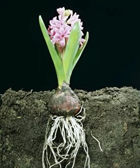
Hyacinth LA 102 Cross Section in earth showing roots © J. M. Labat ARDEA LONDONLA-102 HYACINTH - DEVELOPMENT STAGE 4 Cross Section in earth showing roots Jean Michel Labat Please note that prints are for personal display purposes only and may not be reproduced in anyway
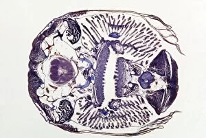
Microscopic Fish Gill - tranverse section through gill region of fish. Fish anatomyJC-429 Microscopic Fish Gill - tranverse section through gill region of fish. Fish anatomy. John Clegg Please note that prints are for personal display purposes only
Light Micrograph (LM): A Tranverse section of a stem of a Common or European Ash tree (Fraxinus excelsior)LRDS-258 Light Micrograph (LM): A Tranverse section of a stem of a Common or European Ash tree Magnification x12 (on 10.5 cm width print) Fraxinus excelsior David Spears (Last Refuge)
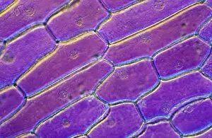
Light Micrograph (LM): Onion skin cells; Magnification x600 (on 10. 5 cm width print)LRDS-252 Light Micrograph (LM): Onion skin cells; Magnification x600 (on 10.5 cm width print) David Spears (Last Refuge) / ardea.com Last Refuge Please note that prints are for personal display
Light Micrograph (LM): Longitudinal section of stem of Crown of Thorns (Euphorbia splendens)LRDS-248 Light Micrograph (LM): Longitudinal section of stem of Crown of Thorns Magnification x1200 (on 10.5 cm width print) Euphorbia splendens David Spears (Last Refuge)
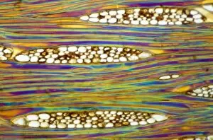
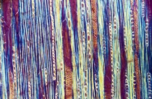
Light Micrograph (LM): Longitudinal section of Mahogani (Swietenia mahagoni); Magnification x600 (on 10)LRDS-246 Light Micrograph (LM): Longitudinal section of Mahogani Magnification x600 (on 10.5 cm width print) Swietenia mahagoni David Spears (Last Refuge)
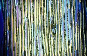
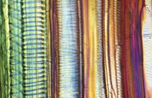
Light Micrograph (LM): Longitudinal section of Scots pine wood, (Pinus sylvestris); Magnification x480 (on 10)LRDS-245 Light Micrograph (LM): Longitudinal section of Scots pine wood, Magnification x480 (on 10.5 cm width print) Pinus sylvestris David Spears (Last Refuge)
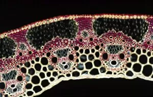
Light Micrograph (LM): A transverse section of a straw of Wheat; Magnification x600 (on 10. 5 cm width print)LRDS-239 Light Micrograph (LM): A transverse section of a straw of Wheat Magnification x600 (on 10.5 cm width print) David Spears (Last Refuge)
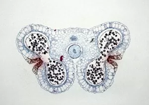
Light Micrograph (LM): Transverse section of Lilium Anthers with Mature Pollen; Magnification x15 (on 10)LRDS-234 Light Micrograph (LM): Transverse section of Lilium Anthers with Mature Pollen Magnification x15 (on 10.5 cm width print) David Spears (Last Refuge)
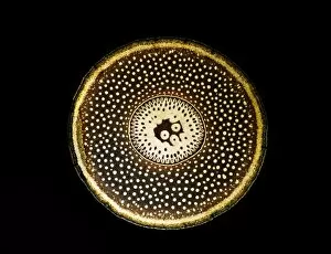
Light Micrograph (LM): A transverse section of a root of Conifer (Pandanus sp. ); Magnification x30 (on 10)LRDS-210 Light Micrograph (LM): A transverse section of a root of Conifer Magnification x30 (on 10.5 cm width print) Pandanus sp
Light Micrograph (LM): A transverse section of an aerial root of Orchid (Dendrobium sp. ); Magnification x30 (on 10)LRDS-209 Light Micrograph (LM): A transverse section of an aerial root of Orchid Magnification x30 (on 10.5 cm width print) Dendrobium sp
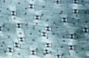
Light Micrograph (LM): A transverse section of a leaf of a Tulip (Tulipa sp)LRDS-202 Light Micrograph (LM): A transverse section of a leaf of a Tulip (Tulipa sp.) showing Stomata Magnification x600 (on 10.5 cm width print) Tulipa sp
Light Micrograph (LM): Longitudinal section of old sunflower stem(Helianthus sp)LRDS-199 Light Micrograph (LM): Longitudinal section of old sunflower stem(Helianthus sp.) showing spiral chloroplasts Magnification x1200 (on 10.5 cm width print)
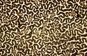
Light Micrograph (LM): A concentration of cells on the epidermis of a plant showing stomataLRDS-197 Light Micrograph (LM): A concentration of cells on the epidermis of a plant showing stomata David Spears (Last Refuge)
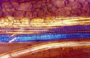
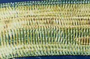
Light Micrograph (LM): Longitudinal section shows Scalariform vessels of Fern; Magnification x1200 (on 10)LRDS-196 Light Micrograph (LM): Longitudinal section shows Scalariform vessels of Fern Magnification x1200 (on 10.5 cm width print) David Spears (Last Refuge)
Light Micrograph (LM): A longitudinal section f a Ribes sp. stem; Magnification x600 (on 10. 5 cm width print)LRDS-194 Light Micrograph (LM): A longitudinal section of a Ribes sp. stem Magnification x600 (on 10.5 cm width print) Ribes sp