mail_outline sales@mediastorehouse.com
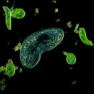
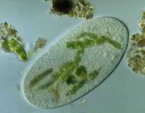
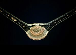
Light Micrograph (LM): Protozoans: Kidney shaped ciliate surrounded by Euglena spLRDS-316 Light Micrograph (LM): Protozoans: Kidney shaped ciliate surrounded by Euglena sp. Magnification x 900 (when printed A4, 29.7 cm wide) Tillina sp
Light Micrograph (LM): A transverse section of a stem of Whisk Fern (Psilotum nudum); Magnification x18 (on 10)LRDS-167 Light Micrograph (LM): A transverse section of a stem of Whisk Fern (Psilotum nudum) Magnification x18 (on 10.5 cm width print) Psilotum nudum David Spears (Last Refuge)
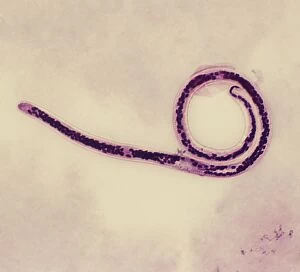
Light Micrograph (SEM): Micro-filarial worm - Magnification x 3000 (if print A4 size: 29. 7 cm wide)LRDS-314 Light Micrograph (SEM): Micro-filarial worm Magnification x 3000 (if print A4 size: 29.7 cm wide) Wuchereria bancrofti Elephantiasis David Spears (Last Refuge)
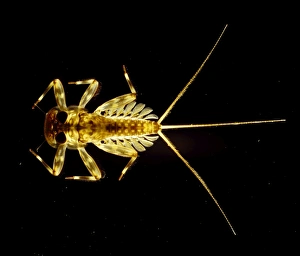
Light Micrograph : Flattened Mayfly Nymph, Magnification x 10 (A4 size: 29. 7 cm width)LRDS-80 Flattened Mayfly Nymph Light Micrograph Ecdyonurus venosus Magnification x 10 (A4 size: 29.7 cm width) Credit: David Spears (last refuge)
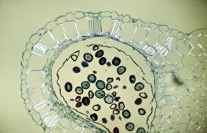
Light Micrograph (LM): Transverse section of Dehiscence Lilium Anthers with Pollen; Magnification x300 (on 10)LRDS-200 Light Micrograph (LM): Transverse section of Dehiscence Lilium Anthers with Pollen Magnification x300 (on 10.5 cm width print) David Spears (Last Refuge)
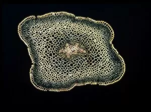
Light Micrograph: Ciliate - Magnification x 750 (when printed A4, 29. 7 cm wide)LRDS-315 Light Micrograph: Ciliate Magnification x 750 (when printed A4, 29.7 cm wide) Frontonia sp. David Spears (Last Refuge) / ardea.com Last Refuge contact details: prints@ardea.com tel
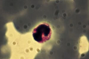
Light Micrograph: Plasmodium: a parasitic protozoa in blood; Magnification x 7, 500 (if print A4 size: 29. 7 cm wide)LRDS-313 Light Micrograph: Plasmodium: a parasitic protozoa in blood; Magnification x 7, 500 (if print A4 size: 29.7 cm wide) David Spears (Last Refuge) / ardea.com Last Refuge contact details
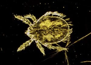
Dark Field Light Micrograph: Scrub Typhus Mite Chigger - Magnification x 125 (if print A4 size: 29. 7 cm wide)LRDS-309 Dark Field Light Micrograph: Scrub Typhus Mite Chigger Magnification x 125 (if print A4 size: 29.7 cm wide) Leptotrombidium akamushi David Spears (Last Refuge)
Light Micrograph (LM): Filaments of fungi Endotrophic Mycorrhiza live within cells of a root; Magnification x600LRDS-254 Light Micrograph (LM): Filaments of fungi Endotrophic Mycorrhiza live within cells of a root Magnification x600 (on 10.5 cm width print) David Spears (Last Refuge)
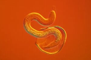
Light Micrograph - a roundworm hatching from its egg against an orange background. CHI0515LRDS-162 Light Micrograph - a roundworm hatching from its egg against an orange background. David Spears (Last Refuge) / ardea.com Last Refuge contact details: prints@ardea.com tel
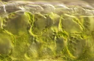
Light Micrograph (LM): plant cell chloroplasts - the site where photosynthesis takes placeLRDS-192 Light Micrograph (LM): plant cell chloroplasts - the site where photosynthesis takes place David Spears (Last Refuge)
Light Micrograph (LM): Cells from a hair on the stamen of the common spiderwort (Tradescantia)LRDS-171 Light Micrograph (LM): Cells from a hair on the stamen of the common spiderwort (Tradescantia) Tradescantia sp David Spears (Last Refuge)
Light Micrograph (LM): A transverse section of a Fig leaf); Magnification x15 (on 10. 5 cm width print)LRDS-187 Light Micrograph (LM): A transverse section of a Fig leaf) Magnification x15 (on 10.5 cm width print) David Spears (Last Refuge)
Light Micrograph (LM): The cellular struture of a liverwort plant (Hepatica); Magnification x1200 (on 10)LRDS-190 Light Micrograph (LM): The cellular struture of a liverwort plant (Hepatica) Magnification x1200 (on 10.5 cm width print) Hepatica sp David Spears (Last Refuge)
Light Micrograph (LM): A single cell from a hair on the stamen of the common spiderwort (Tradescantia)LRDS-170 Light Micrograph (LM): A single cell from a hair on the stamen of the common spiderwort (Tradescantia) Tradescantia sp David Spears (Last Refuge)
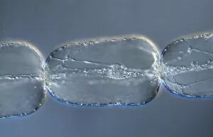
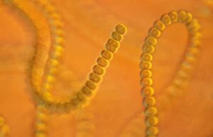
Light Micrograph: Cyanobacterium. ; Magnification x 3, 000 (A4 size: 29. 7 cm width)LRDS-7 Cyanobacterium Light Micrograph Anabaena sp Magnification x 3, 000 (A4 size: 29.7 cm width) Credit: David Spears (last refuge)
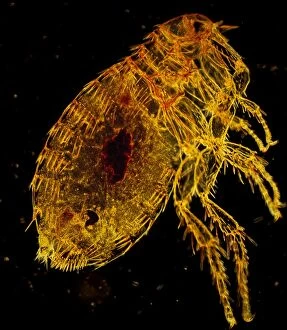
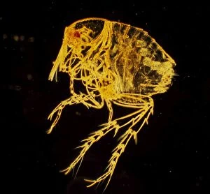
Oriental Rat Flea, Magnification x 200 (A4 size: 29. 7 cm width)LRDS-53 Oriental Rat Flea Dark Field Light Micrograph (LM) Xenopsylla cheopis Magnification x 200 (A4 size: 29.7 cm width) Credit
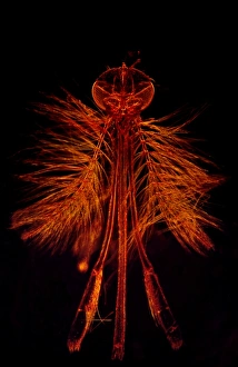
Dark Field Light Micrograph: Mosquito - male. ; Magnification x 65 (A4 size: 29. 7 cm width)LRDS-4 Mosquito - male Dark Field Light Micrograph (LM) Anopheles sp Magnification x 65 (A4 size: 29.7 cm width) Credit: David Spears (last refuge)
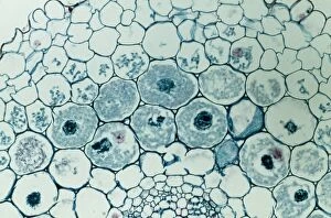
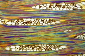
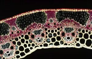
Light Micrograph (LM): A Tranverse section of a stem of a Common or European Ash tree (Fraxinus excelsior)LRDS-258 Light Micrograph (LM): A Tranverse section of a stem of a Common or European Ash tree Magnification x12 (on 10.5 cm width print) Fraxinus excelsior David Spears (Last Refuge)
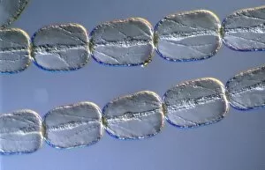
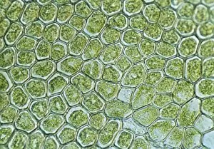
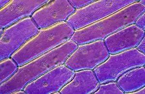
Light Micrograph (LM): Onion skin cells; Magnification x600 (on 10. 5 cm width print)LRDS-252 Light Micrograph (LM): Onion skin cells; Magnification x600 (on 10.5 cm width print) David Spears (Last Refuge) / ardea.com Last Refuge Please note that prints are for personal display
Dark Field Light Micrograph: Chigoe Flea; Magnification x 200 (A4 size: 29. 7 cm width)LRDS-25 Chigoe Flea Dark Field Light Micrograph (LM) Tunga penetrans Magnification x 200 (A4 size: 29.7 cm width) Credit: David Spears (last refuge)
Light Micrograph (LM): Longitudinal section of stem of Crown of Thorns (Euphorbia splendens)LRDS-248 Light Micrograph (LM): Longitudinal section of stem of Crown of Thorns Magnification x1200 (on 10.5 cm width print) Euphorbia splendens David Spears (Last Refuge)
Light Micrograph (LM): Longitudinal section of Mahogani (Swietenia mahagoni); Magnification x600 (on 10)LRDS-246 Light Micrograph (LM): Longitudinal section of Mahogani Magnification x600 (on 10.5 cm width print) Swietenia mahagoni David Spears (Last Refuge)
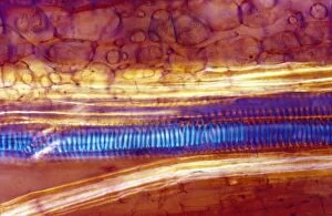
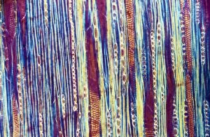
Light Micrograph (LM): Longitudinal section of Scots pine wood, (Pinus sylvestris); Magnification x480 (on 10)LRDS-245 Light Micrograph (LM): Longitudinal section of Scots pine wood, Magnification x480 (on 10.5 cm width print) Pinus sylvestris David Spears (Last Refuge)
Light Micrograph (LM): A transverse section of a straw of Wheat; Magnification x600 (on 10. 5 cm width print)LRDS-239 Light Micrograph (LM): A transverse section of a straw of Wheat Magnification x600 (on 10.5 cm width print) David Spears (Last Refuge)
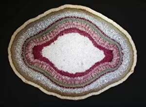
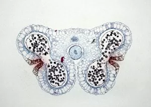
Light Micrograph (LM): Transverse section of Lilium Anthers with Mature Pollen; Magnification x15 (on 10)LRDS-234 Light Micrograph (LM): Transverse section of Lilium Anthers with Mature Pollen Magnification x15 (on 10.5 cm width print) David Spears (Last Refuge)
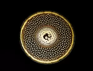
Light Micrograph (LM): A transverse section of a root of Conifer (Pandanus sp. ); Magnification x30 (on 10)LRDS-210 Light Micrograph (LM): A transverse section of a root of Conifer Magnification x30 (on 10.5 cm width print) Pandanus sp
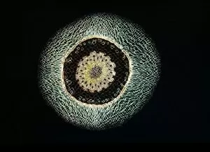
Light Micrograph (LM): A transverse section of an aerial root of Orchid (Dendrobium sp. ); Magnification x30 (on 10)LRDS-209 Light Micrograph (LM): A transverse section of an aerial root of Orchid Magnification x30 (on 10.5 cm width print) Dendrobium sp
Light Micrograph (LM): A transverse section of a leaf of a Tulip (Tulipa sp)LRDS-202 Light Micrograph (LM): A transverse section of a leaf of a Tulip (Tulipa sp.) showing Stomata Magnification x600 (on 10.5 cm width print) Tulipa sp
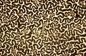
Light Micrograph (LM): Longitudinal section of old sunflower stem(Helianthus sp)LRDS-199 Light Micrograph (LM): Longitudinal section of old sunflower stem(Helianthus sp.) showing spiral chloroplasts Magnification x1200 (on 10.5 cm width print)
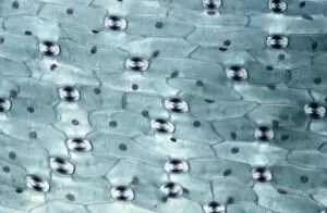
Light Micrograph (LM): A concentration of cells on the epidermis of a plant showing stomataLRDS-197 Light Micrograph (LM): A concentration of cells on the epidermis of a plant showing stomata David Spears (Last Refuge)
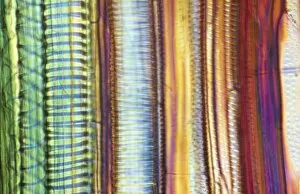
Light Micrograph (LM): Longitudinal section shows Scalariform vessels of Fern; Magnification x1200 (on 10)LRDS-196 Light Micrograph (LM): Longitudinal section shows Scalariform vessels of Fern Magnification x1200 (on 10.5 cm width print) David Spears (Last Refuge)
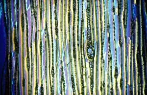
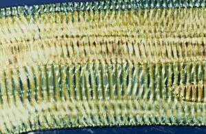
Light Micrograph (LM): A longitudinal section f a Ribes sp. stem; Magnification x600 (on 10. 5 cm width print)LRDS-194 Light Micrograph (LM): A longitudinal section of a Ribes sp. stem Magnification x600 (on 10.5 cm width print) Ribes sp