mail_outline sales@mediastorehouse.com
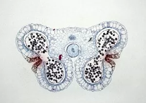
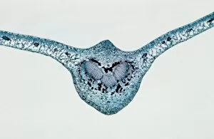
Light Micrograph (LM): Transverse section of Lilium Anthers with Mature Pollen; Magnification x15 (on 10)LRDS-234 Light Micrograph (LM): Transverse section of Lilium Anthers with Mature Pollen Magnification x15 (on 10.5 cm width print) David Spears (Last Refuge)
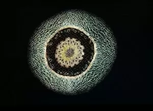
Light Micrograph (LM): A transverse section of a root of Conifer (Pandanus sp. ); Magnification x30 (on 10)LRDS-210 Light Micrograph (LM): A transverse section of a root of Conifer Magnification x30 (on 10.5 cm width print) Pandanus sp
Light Micrograph (LM): A transverse section of an aerial root of Orchid (Dendrobium sp. ); Magnification x30 (on 10)LRDS-209 Light Micrograph (LM): A transverse section of an aerial root of Orchid Magnification x30 (on 10.5 cm width print) Dendrobium sp
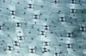
Light Micrograph (LM): A transverse section of a leaf of a Tulip (Tulipa sp)LRDS-202 Light Micrograph (LM): A transverse section of a leaf of a Tulip (Tulipa sp.) showing Stomata Magnification x600 (on 10.5 cm width print) Tulipa sp
Light Micrograph (LM): Longitudinal section of old sunflower stem(Helianthus sp)LRDS-199 Light Micrograph (LM): Longitudinal section of old sunflower stem(Helianthus sp.) showing spiral chloroplasts Magnification x1200 (on 10.5 cm width print)
Light Micrograph (LM): A concentration of cells on the epidermis of a plant showing stomataLRDS-197 Light Micrograph (LM): A concentration of cells on the epidermis of a plant showing stomata David Spears (Last Refuge)
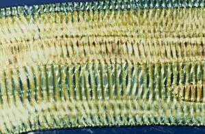
Light Micrograph (LM): Longitudinal section shows Scalariform vessels of Fern; Magnification x1200 (on 10)LRDS-196 Light Micrograph (LM): Longitudinal section shows Scalariform vessels of Fern Magnification x1200 (on 10.5 cm width print) David Spears (Last Refuge)
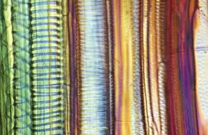
Light Micrograph (LM): A longitudinal section f a Ribes sp. stem; Magnification x600 (on 10. 5 cm width print)LRDS-194 Light Micrograph (LM): A longitudinal section of a Ribes sp. stem Magnification x600 (on 10.5 cm width print) Ribes sp
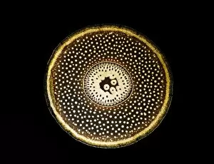
Light Micrograph (LM): transverse section of Prunus Leaf; Magnification x18 (on 10. 5 cm width print)LRDS-181 Light Micrograph (LM): transverse section of Prunus Leaf Magnification x18 (on 10.5 cm width print) Prunus sp David Spears (Last Refuge)
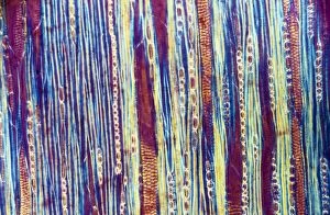
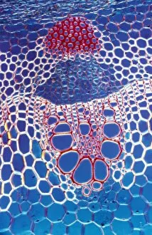
Light Micrograph (LM): A transverse section shows Vascular Bundle in Helianthus stem; Magnification x600 (on 10)LRDS-177 Light Micrograph (LM): A transverse section shows Vascular Bundle in Helianthus stem Magnification x600 (on 10.5 cm width print) Helianthus sp David Spears (Last Refuge)
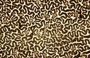
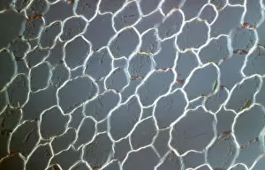
Light Micrograph (LM): transvers section shows Parenchyma Ground Tissue in Buttercup stem; Magnification x600 (on 10)LRDS-176 Light Micrograph (LM): transvers section shows Parenchyma Ground Tissue in Buttercup stem Magnification x600 (on 10.5 cm width print) David Spears (Last Refuge)
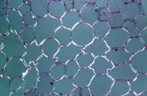
Light Micrograph (LM): tranverse section shows Parenchyma or packing tissue from Cucurbita Stem; Magnification x600LRDS-174 Light Micrograph (LM): tranverse section shows Parenchyma or packing tissue from Cucurbita Stem Magnification x600 (on 10.5 cm width print) David Spears (Last Refuge)
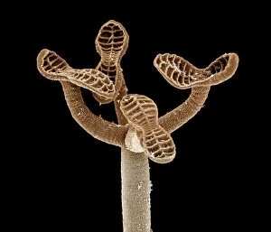
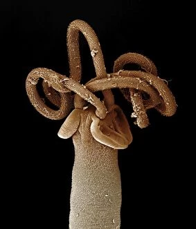
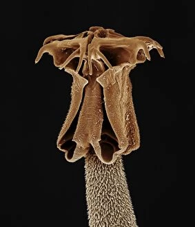
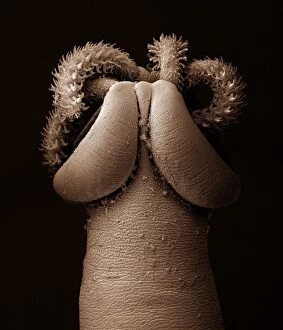
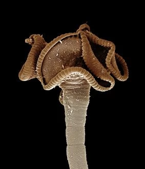
Tapeworm from shark CHI0876LRDS-161 Tapeworm from shark David Spears (Last Refuge) / ardea.com Last Refuge Please note that prints are for personal display purposes only and may not be reproduced in any way
Tapeworm from shark CHI0882LRDS-160 Tapeworm from shark David Spears (Last Refuge) / ardea.com Last Refuge Please note that prints are for personal display purposes only and may not be reproduced in any way
Tapeworm from shark CHI0874LRDS-159 Tapeworm from shark David Spears (Last Refuge) / ardea.com Last Refuge Please note that prints are for personal display purposes only and may not be reproduced in any way
Tapeworm from shark CHI0868LRDS-156 Tapeworm from shark David Spears (Last Refuge) / ardea.com Last Refuge Please note that prints are for personal display purposes only and may not be reproduced in any way
Tapeworm from shark CHI0884LRDS-155 Tapeworm from shark David Spears (Last Refuge) / ardea.com Last Refuge Please note that prints are for personal display purposes only and may not be reproduced in any way
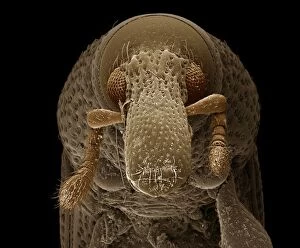
Scanning Electron Micrograph: Woodboring Weevil, Magnification x 222 (A4 size: 29. 7 cm width)LRDS-147 Woodboring Weevil Scanning Electron Micrograph Euophryum confine Magnification x 222 (A4 size: 29.7 cm width) Coloured by hand to enhance natural features
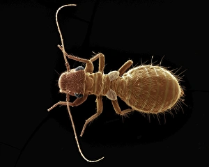
Scanning Electron Micrograph (SEM): Termite, Magnification x 30 (A4 size: 29. 7 cm width)LRDS-145 Termite Scanning Electron Micrograph (SEM) Reticulitermes lucifugus Magnification x 30 (A4 size: 29.7 cm width) Coloured by hand to enhance natural features
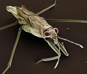
Scanning Electron Micrograph (SEM): Pond Skater, Magnification x 40 (A4 size: 29. 7 cm width)LRDS-111 Pond Skater Scanning Electron Micrograph (SEM) Gerris lacustris Magnification x 40 (A4 size: 29.7 cm width) Coloured by hand to enhance natural features
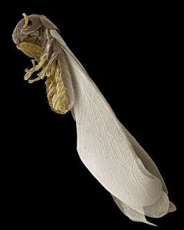
Scanning Electron Micrograph (SEM): Psocid, Magnification x 100 (A4 size: 29. 7 cm width)LRDS-104 Psocid Scanning Electron Micrograph (SEM) Lepinotus sp. Magnification x 100 (A4 size: 29.7 cm width) Coloured by hand to enhance natural features
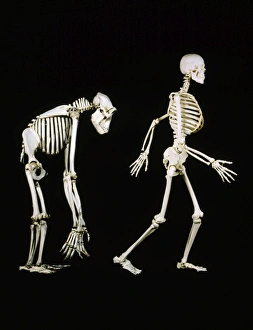
Skeleton - Gorilla & ManLA-78 SKELETONS - HUMAN & GORILLA Man walking upright Jean Michel Labat Please note that prints are for personal display purposes only and may not be reproduced in any way
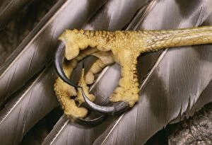
Sparrow-Hawk Close up of talonsJLMO-3453 SPARROWHAWK - close-up of talons Accipitor nisus John Mason Please note that prints are for personal display purposes only and may not be reproduced in any way
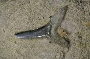
Shark's Tooth Fossil London clay, Herne Bay, Kent, UKJLMO-2595 Fossil - Shark's Tooth Fossil London clay, Herne Bay, Kent, UK. Striatolamna sp. John Mason Please note that prints are for personal display purposes only
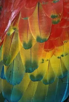
Scarlet Macaw - FeathersAW-3594 Scarlet Macaw - Feathers Ara macao Adrian Warren Please note that prints are for personal display purposes only and may not be reproduced in any way
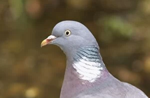
Wood Pigeon CK 1988 Columba palumbus © Chris knights / ARDEA LONDONCK-1988 WOODPIGEON - close-up of head Columba palumbus Chris Knights Please note that prints are for personal display purposes only and may not be reproduced in any way
Tadpoles – common frog – 4 weeks old – 2 x at 35mm Bedfordshire UK 003915hBB-1243 Tadpoles of common frog - 4 weeks old, 2 x at 35mm Bedfordshire, UK Rana temporaria Brian Bevan Please note that prints are for personal display purposes only
Tadpoles – common frog – 4 weeks old – 1 x at 35mm – black background Bedfordshire UK 003848BB-1241 Tadpoles of common frog - 4 weeks old, 1 x at 35mm Bedfordshire, UK Rana temporaria Brian Bevan Please note that prints are for personal display purposes only
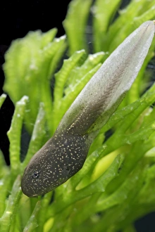
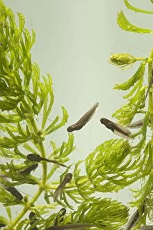
Tadpoles – common frog – 9 days old – 0. 5 x at 35mm – green background Bedfordshire UK 003729BB-1233 9 days old Tadpoles of common frog - 0.5 x at 35mm Bedfordshire, UK Rana temporaria Brian Bevan Please note that prints are for personal display purposes only
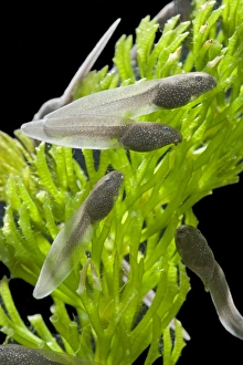
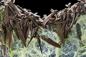
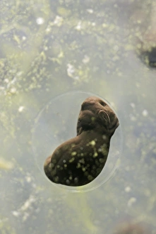
Tadpoles – common frog – 9 days old – 2 x at 35mm – black background Bedfordshire UK 003667hBB-1231 9 days old Tadpoles of common frog - 2 x at 35mm Bedfordshire, UK Rana temporaria Brian Bevan Please note that prints are for personal display purposes only
Tadpoles – common frog – 9 days old – 1 x at 35mm – black background Bedfordshire UK 003646BB-1230 9 days old Tadpoles of common frog - 1 x at 35mm Bedfordshire, UK Rana temporaria Brian Bevan Please note that prints are for personal display purposes only
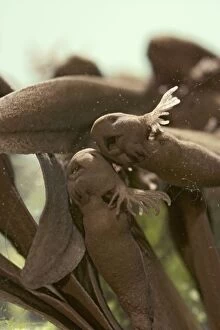
Tadpoles – emerging - common frog – 4 x at 35mm – green background Bedfordshire UK 003592BB-1223 Common frog Tadpoles - emerging, 4 x at 35mm Bedfordshire, UK Rana temporaria Brian Bevan Please note that prints are for personal display purposes only and may not be reproduced in any way
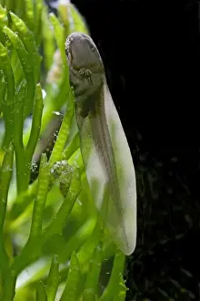
Tadpoles – emerging - common frog – 1 x at 35mm – black background Bedfordshire UK 003587BB-1222 Common frog Tadpoles - emerging, 1 x at 35mm Bedfordshire, UK Rana temporaria Brian Bevan Please note that prints are for personal display purposes only and may not be reproduced in any way
Frogspawn – 7 day old - common frog – 4 x at 35mm – green background Bedfordshire UK 003581hBB-1220 7 day old Frogspawn of common frog - 4 x at 35mm Bedfordshire, UK Rana temporaria Brian Bevan Please note that prints are for personal display purposes only
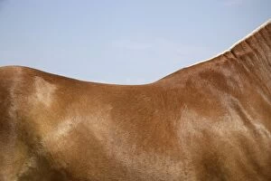
Horse. BackJD-19366 Horse - Back John Daniels Please note that prints are for personal display purposes only and may not be reproduced in any way. contact details: prints@ardea.com tel: + 44 (0) 20 8318 1401
House Spider Showing eyes palps and jawsFEU-166 House Spider - Showing eyes palps and jaws Tegenaria domestica Geoff du Feu Please note that prints are for personal display purposes only and may not be reproduced in any way
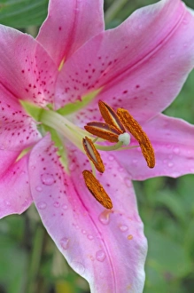
Flower head of Lilium "Sorbonne" - An interesting cultivar of the bulbous perennial splashed with spotsDAD-1934 Flower head of Lilium "Sorbonne" - An interesting cultivar of the bulbous perennial splashed with spots and contrasting pollen-bearing anthers East Sussex garden in July
Torch Cactus (Hybrid) - Trichocereus - Arizona-not native to Arizona- under cultivationCAN-3143 Torch Cactus (Hybrid) - not native to Arizona, under cultivation Arizona, USA Trichocereus John Cancalosi Please note that prints are for personal display purposes only
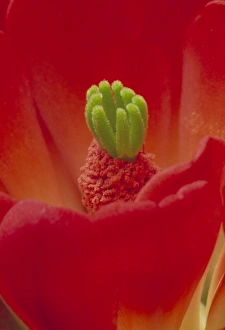
Claret Cup Cactus Blossum - New Mexico, USACAN-2719 Claret Cup Cactus Blossom New Mexico, USA John Cancalosi Please note that prints are for personal display purposes only and may not be reproduced in any way
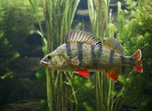
Perch – use weeds as camouflage – dorsal fin up – side view - Europe UKBB-933 Perch - use weeds as camouflage, dorsal fin up, turning UK Perca fluviatilis Brian Bevan Please note that prints are for personal display purposes only and may not be reproduced in any way
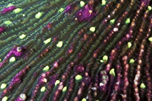
Mushroom Coral - with bright green tentacular lobesAU-35-MS Mushroom Coral - with bright green tentacular lobes Australia Fungia scutaria Auscape Please note that prints are for personal display purposes only and may not be reproduced in any way
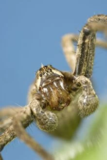
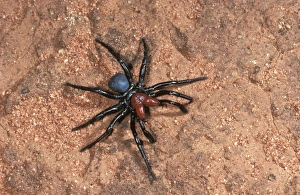
Red-headed mouse spider male searching for mate. Only the male is called ‘red-headed'. Australia CAH00097AU-3-cah Red-headed mouse SPIDER - male searching for mate Australia Missulena insigne Only the male is called red-headed
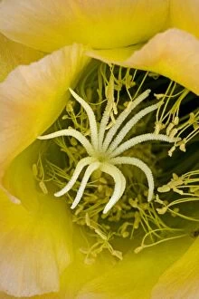
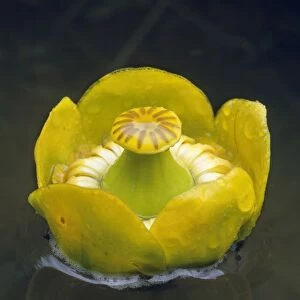
Yellow WaterlilyAL-2101 Yellow Waterlily Nuphar lutea Ake Lindau Please note that prints are for personal display purposes only and may not be reproduced in any way
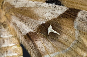
Tau emperor - Close-up of Wings of femaleVG-303 Tau emperor - Close-up of Wings of female Aglia tau Paul Van Gaalen Please note that prints are for personal display purposes only and may not be reproduced in any way
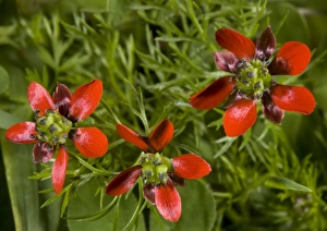
Pheasant's eye - cornfield weed, very rare in UKROG-11706 Pheasant's eye - an arable, wheat / cornfield weed, Greece. very rare in UK. Adonis annua Bob Gibbons Please note that prints are for personal display purposes only
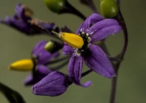
Bittersweet NightshadeROG-11475 Bittersweet Nightshade UK Solanum dulcamara Bob Gibbons Please note that prints are for personal display purposes only and may not be reproduced in any way
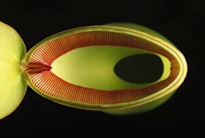
Pitcher Plant - from abovePPG-1679 Pitcher Plant - from above Nepenthes ventricosa Pascal Goetgheluck Please note that prints are for personal display purposes only and may not be reproduced in any way
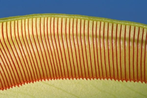
Pitcher Plant - the top of the pitcher are covered with downward facing hairs meant to prevent the preys from going upPPG-1645 Pitcher Plant - the top of the pitcher are covered with downward facing hairs meant to prevent their prey from going up Nepenthes ventricosa Pascal Goetgheluck Please note that prints are