mail_outline sales@mediastorehouse.com
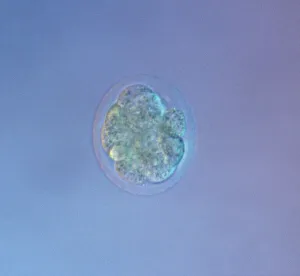
Picture No. 11675528Mouse Embryo at Eight Cell Stage. Date:
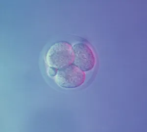
Picture No. 11675527Mouse Embryo at Four Cell Stage & a Polar Body. Date:
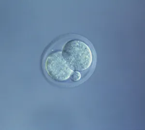
Picture No. 11675526Mouse Embryo at Two Cell Stage & a Polar Body. Date:
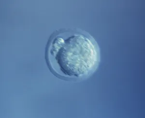
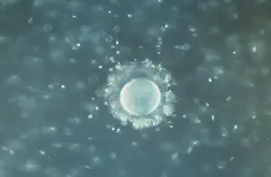
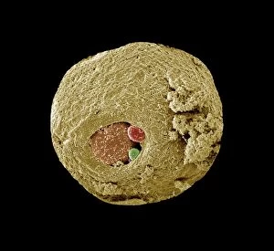
Picture No. 11675525Mouse Embryo at Single Cell Stage and Polar Body. Date:
Picture No. 11675522A human Ovum surrounded by Sperm - being fertilized. Date:
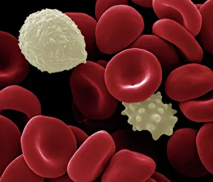
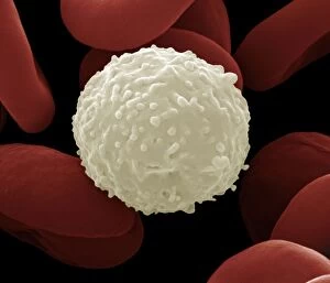
Picture No. 11675612Scanning Electron Micrograph (SEM): Human White and Red Blood Cells. Date:
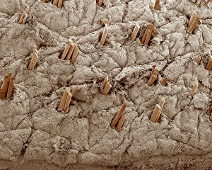
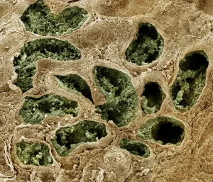
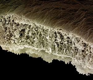
Picture No. 10876997Scanning Electron Micrograph (SEM): Human Skin with Hair Follicles Date:
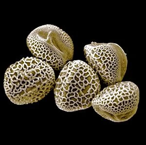
Picture No. 11675582Scanning Electron micrograph (SEM): Lily Pollen grains. Date:
Picture No. 11675585Scanning Electron micrograph (SEM)showing stomata on a Yew Leaf. Date:
Picture No. 10873704Sleeping Sickness Parasite in red blood cells (Trypanosoma sp. ) Date:
DDE-90029660Photo Cell window blinds at Instute du Monde Arabe, Arab World Institute, Paris, France. Date: 30/01/2009
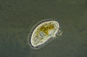
Picture No. 11014599Freshwater Ciliates common in pools and ponds consisting of a single cell and feed by ingesting other tiny organisms such as bacteria and other protozoa Date:
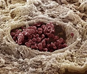
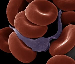
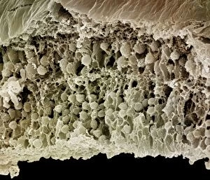
Picture No. 10876987Scanning Electron Micrograph (SEM): Human skin section across vein showing red blood cells Date:
Picture No. 11675577Scanning Electron Micrograph (SEM): Human testis tubes - Sperm cells forming in seminal tubes of testis. Date:
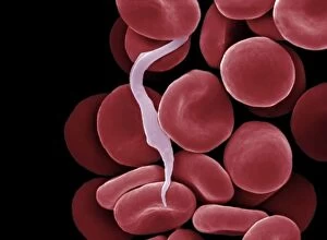
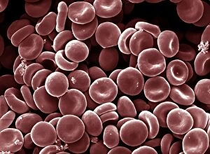
Picture No. 10873675Human White and Red Blood Cells Date:
Picture No. 10873703Sleeping Sickness Parasite in red blood cells (Trypanosoma sp.) Date:
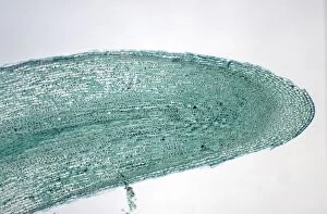
Picture No. 10852165Broad Bean Root Tip - shows cell division / mitosis (Vicia faba) Date:
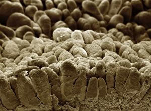
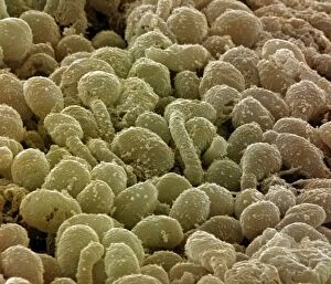
Picture No. 10877012Scanning Electron Micrograph (SEM): human intestine showing villi Date:
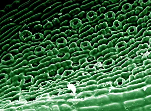
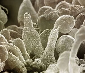
Picture No. 10876996Scanning Electron Micrograph (SEM): Human Retina showing photoreceptors rod and cone cells Date:
Picture No. 10876993Scanning Electron Micrograph (SEM): Human Retina showing photoreceptors rod and cone cells Date:
Picture No. 10876995Scanning Electron Micrograph (SEM): Human Retina showing photoreceptors rod and cone cells Date:
Picture No. 10876994Scanning Electron Micrograph (SEM): Human Retina showing photoreceptors rod and cone cells Date:
Picture No. 10876988Scanning Electron Micrograph (SEM): Early human embryo, one cell removed for genetic analysis Date:
Picture No. 10876986Scanning Electron Micrograph (SEM): Human Red Blood Cells Date: