mail_outline sales@mediastorehouse.com
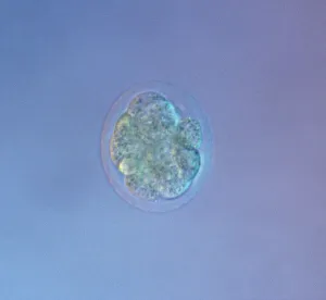
Picture No. 11675528Mouse Embryo at Eight Cell Stage. Date:
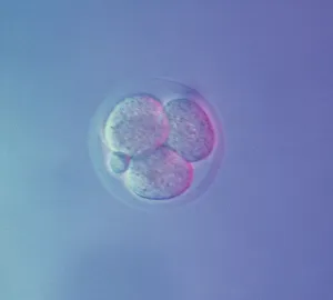
Picture No. 11675527Mouse Embryo at Four Cell Stage & a Polar Body. Date:
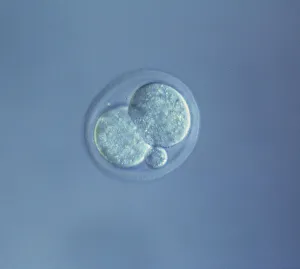
Picture No. 11675526Mouse Embryo at Two Cell Stage & a Polar Body. Date:
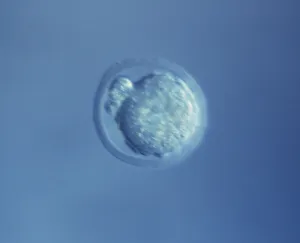
Picture No. 11675525Mouse Embryo at Single Cell Stage and Polar Body. Date:
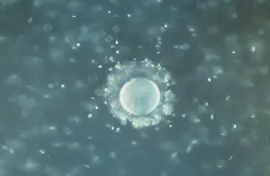
Picture No. 11675522A human Ovum surrounded by Sperm - being fertilized. Date:
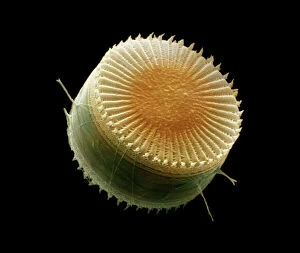
Picture No. 11675478Scanning Electron Micrograph (SEM): Freshwater Diatom, Cyclotella meneghiniana. Date:
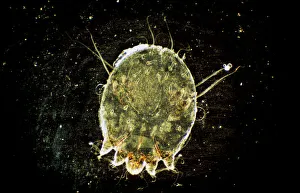
Picture No. 11675613Dark Field Light Micrograph: Scabies Mite. Date:
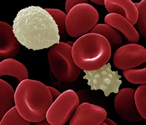
Picture No. 11675612Scanning Electron Micrograph (SEM): Human White and Red Blood Cells. Date:
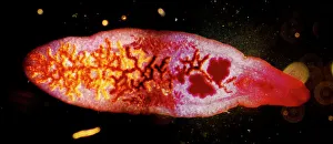
Picture No. 11675599Dark Field Light Micrograph: Lancet Liver Fluke. Date:
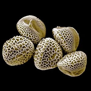
Picture No. 11675582Scanning Electron micrograph (SEM): Lily Pollen grains. Date:
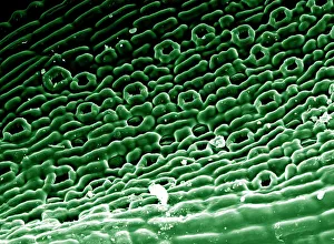
Picture No. 11675585Scanning Electron micrograph (SEM)showing stomata on a Yew Leaf. Date:
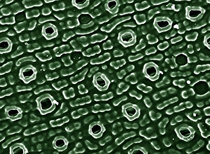
Picture No. 11675628Scanning Electron Micrograph (SEM): Stomata of Yew Leaf. Date:
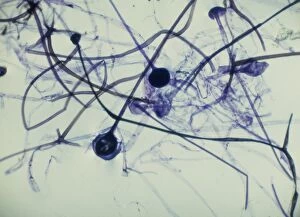
Picture No. 11675563Light Micrograph (LM): Rhizopus sporangia. Date:
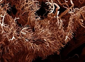
Picture No. 11675581Scanning Electron Micrograph (SEM): a corrosion cast of a gut tumour - Liquid plastic is injected into the blood vessels; it solidifies
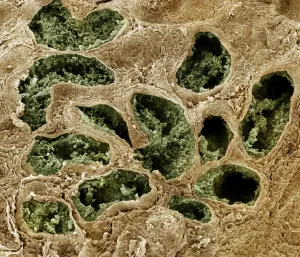
Picture No. 11675577Scanning Electron Micrograph (SEM): Human testis tubes - Sperm cells forming in seminal tubes of testis. Date: