mail_outline sales@mediastorehouse.com
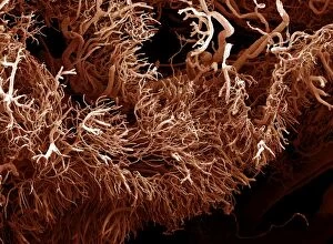
Picture No. 11675581Scanning Electron Micrograph (SEM): a corrosion cast of a gut tumour - Liquid plastic is injected into the blood vessels; it solidifies
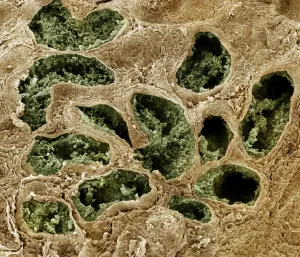
Picture No. 11675577Scanning Electron Micrograph (SEM): Human testis tubes - Sperm cells forming in seminal tubes of testis. Date:
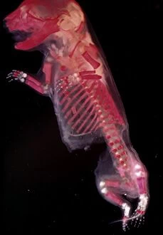
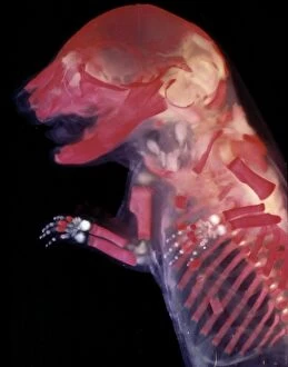
Picture No. 11675517Mouse Embryo - red dye to show bones. Date:
Picture No. 11675516Mouse Embryo - red dye to show bones. Date:
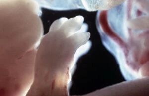
Picture No. 11675515Rat Embryo - The paws of a 17.5 day old rat embryo. Date:
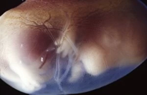
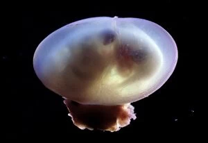
Picture No. 11675514Rat Embryo 17.5 days into its gestation period, in sac with placenta. Date:
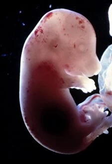
Picture No. 11675513Rat Embryo without its yolk sac, at 17.5 days. Date:
Picture No. 11675512Rat Embryo without its yolk sac, at 17.5 days. Date:
Picture No. 11675511Rat Embryo without its yolk sac, at 17.5 days. Date:
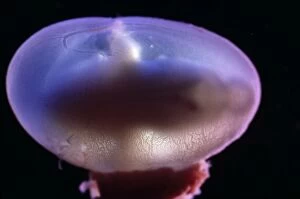
Picture No. 11675510Rat Embryo - 15.5 days into its gestation period - in sac with placenta. Date:
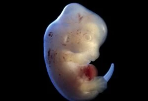
Picture No. 11675509Rat Embryo at 15.5 days, no sac, no placenta. Date:
Picture No. 11675508Rat Embryo - at 15.5 days, no sac, no placenta. Date:
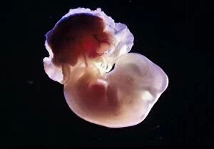
Picture No. 11675506Rat Embryo - 14.5 days into its gestation period - in sac with placenta. Date:
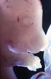
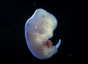
Picture No. 11675504Rat Embryo 13.2 days after fertilisation, out of its yolk sac. Date:
Picture No. 11675505Rat Embryo - 14.5 days into its gestation period - in sac with placenta. Date:
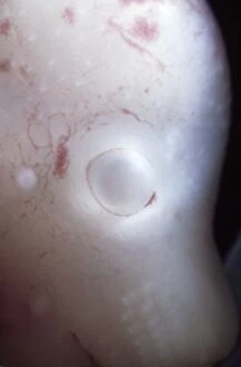
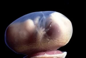
Picture No. 11675502Eye detail of a rat embryo at 13.2 days after fertilisation. Date: