mail_outline sales@mediastorehouse.com
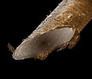
Picture No. 10877003Scanning Electron Micrograph (SEM): Human hair - African Date:
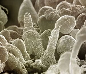
Picture No. 10876996Scanning Electron Micrograph (SEM): Human Retina showing photoreceptors rod and cone cells Date:
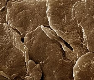
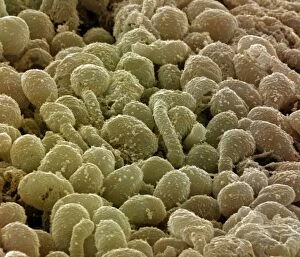
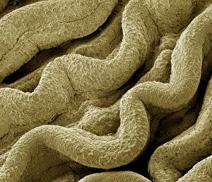
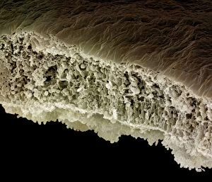
Picture No. 10877001Scanning Electron Micrograph (SEM): Human Skin Date:
Picture No. 10876999Scanning Electron Micrograph (SEM): Human Skin Date:
Picture No. 10876991Scanning Electron Micrograph (SEM): Human testis tubes - Sperm cells forming in seminal tubes of testis Date:
Picture No. 10876993Scanning Electron Micrograph (SEM): Human Retina showing photoreceptors rod and cone cells Date:
Picture No. 10876995Scanning Electron Micrograph (SEM): Human Retina showing photoreceptors rod and cone cells Date:
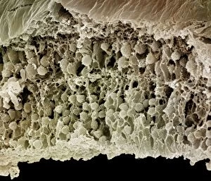
Picture No. 10876989Scanning Electron Micrograph (SEM): Human Fallopian tube Date:
Picture No. 10876994Scanning Electron Micrograph (SEM): Human Retina showing photoreceptors rod and cone cells Date:
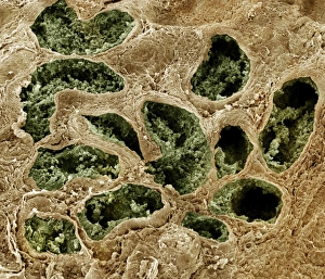
Picture No. 10876992Scanning electron micrograph (SEM) of Kidney glomerulus Date:
Picture No. 10876988Scanning Electron Micrograph (SEM): Early human embryo, one cell removed for genetic analysis Date:
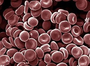
Picture No. 10876986Scanning Electron Micrograph (SEM): Human Red Blood Cells Date:
Picture No. 10874543Scanning Electron Micrograph (SEM): Tapeworm from a teleost fish Date:
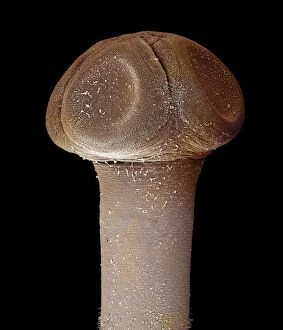
Picture No. 10874544Scanning Electron Micrograph (SEM): human sperm Date:
Picture No. 10895903Tarantula - Urticants hair under SEM (Scanning Electron Microscope) (Lasiodora parahybana) Date:
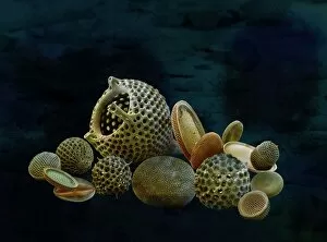
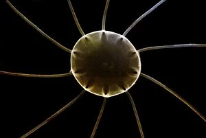
Marine Diatoms and RadiolariansLRMC-17 Scanning Electron Micrograph (SEM): Marine Diatoms and Radiolaria skeletons Magnification x435 (when printed A4, 29.7 cm cm wide)
Scanning Electron Micrograph (SEM): Fusarium, Magnification x 1, 600 (A4 size: 29. 7 cm width)LRDS-60 Fusarium Scanning Electron Micrograph (SEM) Fusarium graminearum Magnification x 1, 600 (A4 size: 29.7 cm width) Coloured by hand to enhance natural features
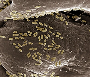
Scanning Electron Micrograph (SEM): E. coli; Magnification x 15, 000 (A4 size: 29. 7 cm width)LRDS-40 E. coli Scanning Electron Micrograph (SEM) Escherichia coli Magnification x 15, 000 (A4 size: 29.7 cm width) Coloured by hand to enhance natural features
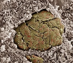
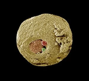
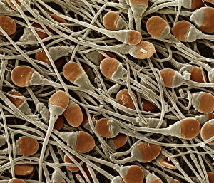
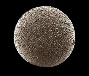
ForaminiferaLRDS-306 Scanning Electron Micrograph (SEM): Foraminifera Magnification x400 (when printed A4 size, 29.7 cm wide) Orbulina universa David Spears (Last Refuge) / ardea.com Last Refuge contact details
Marine DiatomLRDS-293 Scanning Electron Micrograph (SEM): Marine Diatom Magnification x2, 570 (when printed 10.5 cm wide) Bacteriastrum sp David Spears (Last Refuge) / ardea.com Last Refuge contact details
Marine DiatomLRDS-291 Scanning Electron Micrograph (SEM): Marine Diatom Magnification x1, 510 (when printed 10.5 cm wide) David Spears (Last Refuge) / ardea.com Last Refuge contact details: prints@ardea.com tel
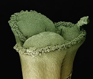
Scanning Electron Micrograph (SEM): stigma of a lily, Magnification x 100 (A4 size: 29. 7 cm width)LRDS-125 stigma of a lily Scanning Electron Micrograph (SEM) Lilium sp. Magnification x 100 (A4 size: 29.7 cm width) Coloured by hand to enhance natural features
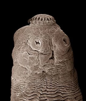
Tapeworm CHI0860LRDS-154 Tapeworm Taenia solium David Spears (Last Refuge) / ardea.com Last Refuge Please note that prints are for personal display purposes only and may not be reproduced in any way
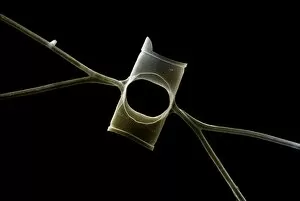
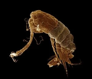
Scanning Electron Micrograph (SEM): Calanus sp. ; Magnification x650 (A4 size: 29. 7 cm width)LRDS-302 Scanning Electron Micrograph (SEM): Calanus sp Magnification x650 (A4 size: 29.7 cm width) David Spears (Last Refuge)
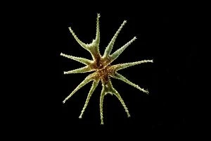
Freshwater DesmidLRDS-303 Scanning Electron Micrograph (SEM): Freshwater Desmid Magnification x260 (when printed 10.5 cm Micrasterias lux David Spears (Last Refuge)