mail_outline sales@mediastorehouse.com
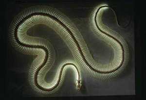
Python Snake Skeleton Light with neon tube bulb, about 15 longKFO-435 Python Snake Skeleton Africa Light with neon tube bulb, about 15 long. Python spp. Kenneth W Fink Please note that prints are for personal display purposes only
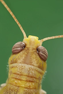
Locust – close up of head top view 003840hBB-1238 Locust - close up of head top view Brian Bevan Please note that prints are for personal display purposes only and may not be reproduced in any way
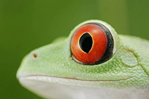
Red-eyed tree frog - close up of eye – South and Central America 003074BB-1099 Red-eyed tree frog - close up of eye South and Central America Agalychnis callidryas Distribution: belize, Costa Rica, Guatemala, Honduras, Mexico
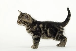
CAT - Tabby KitenLA-1491 CAT - Tabby Kitten, side profile Jean Michel Labat Please note that prints are for personal display purposes only and may not be reproduced in any way
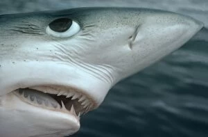
Oceanic Blue Shark Above water, close-up of face showing detail of teeth and eye Eastern PacificVT-8299 Oceanic Blue Shark - Above water, close-up of face showing details of teeth and eye. Also shows ampoules / ampullae of Lorenzini
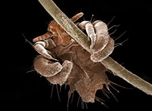
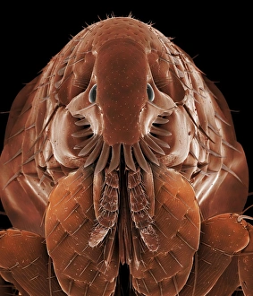
Picture No. 10873599Human Crab Louse (Phthirus pubis) Date:
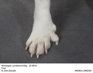
Picture No. 10851731Norwegian Lundehund Dog - Foot Date:
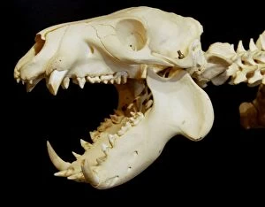
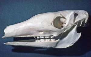
Picture No. 10874838Pigmy Hippo skull (Hexaprotodon liberiensis) Date:
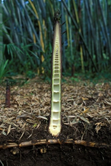
Picture No. 10865930Bamboo - cross section of a shoot emerging from the ground and its rhizome Date:
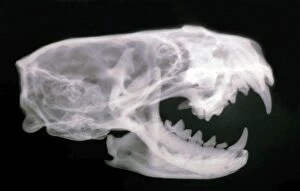
Picture No. 10864939Mink Skull- X ray showing teeth (Mustela vison) Date:
Picture No. 10864934Aardvark - skull showing simple peg like cheek teeth and no incisors or canines (Orycteropus afer) Date:
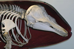
Picture No. 10861874Harbour Porpoise - Skeleton inside shape of skin (Phocoena phocoena) Date:
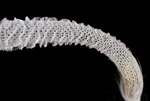
Picture No. 10854313Venus Glass Sponge / Flower Basket (Euplectella) Date:
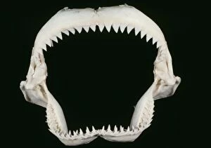
Picture No. 10852952Shark Jaws - skeleton (Carcharhinus sp.) Date:
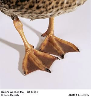
Picture No. 10851777Duck's Webbed feet Date:
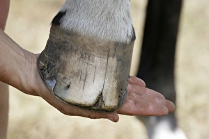
Picture No. 10745632Horse. Split hoof Date:
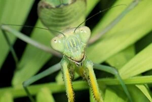
Praying MantisSPH-105 Praying Mantis Sphodromantis lineola Steve Hopkin Please note that prints are for personal display purposes only and may not be reproduced in any way
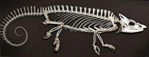
Chameleon - skeletonPM-10696 Chameleon - skeleton Chamaeleo melleri Pat Morris contact details: prints@ardea.com tel: +44 (0) 20 8318 1401
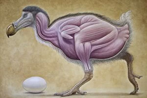
Dodo - muscle studyJH-62 Dodo - muscle study Raphus cucullatus John Holmes contact details: prints@ardea.com tel: +44 (0) 20 8318 1401
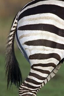
Burchell's / Common / Plains Zebra LA 650 Close up of rear end Equus burchelli © J. M. Labat / ardea. comLA-650 Burchell's / Common / Plains Zebra - Close up of rear end Africa Equus quagga burchelli Jean Michel Labat contact details: prints@ardea.com tel: +44 (0) 20 8318 1401
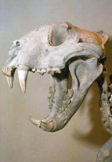
Lion SkullPM-4181 SKELETON - LION SKULL Pat Morris Please note that prints are for personal display purposes only and may not be reproduced in any way. contact details: web@ardea.com tel: + 44 (0) 20 8318 1401
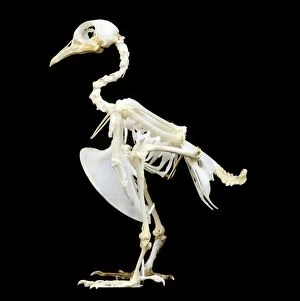
Pigeon SkeletonLA-86 Pigeon Skeleton Jean Michel Labat Please note that prints are for personal display purposes only and may not be reproduced in any way. contact details: web@ardea.com tel: + 44 (0) 20 8318 1401
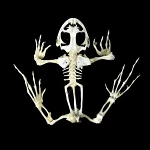
Frog SkeletonLA-84 SKELETON - frog Jean Michel Labat Please note that prints are for personal display purposes only and may not be reproduced in any way. contact details: web@ardea.com tel: + 44 (0) 20 8318 1401
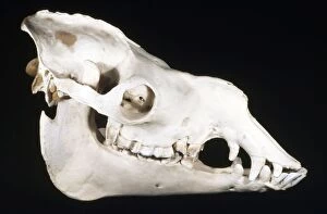
Dromedary Camel Skull - male - dry grasslands - north AfricaKEL-1213 Dromedary Camel Skull - male dry grasslands - north Africa Camelus dromedarius Ken Lucas Please note that prints are for personal display purposes only and may not be reproduced in any way
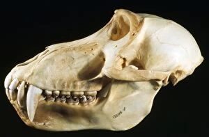
Baboon Skull - male - Eastern Central AfricaKEL-1009 Baboon Skull - male Eastern Central Africa Papio sp. Ken Lucas Please note that prints are for personal display purposes only and may not be reproduced in any way
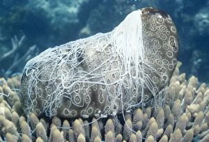
Sea Cucumber - with Cuverian organs / Sticky threads ejected. toxic threads which stick onto a predator & are leftVT-2731 SEA CUCUMBER - with cuvierian organs / sticky threads ejected Family: Holothurian Class: Holothuridaea The Cuvierian organs / sticky threads are part of the digestive tract
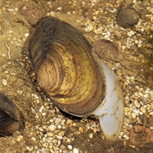
Swan Mussel - Showing threds that attach Mussels to rocksBB-582 Swan MUSSEL - moving, with muscular foot extended Anodonta cygnea showing threads that attach mussel to rocks Brian Bevan Please note that prints are for personal display purposes only
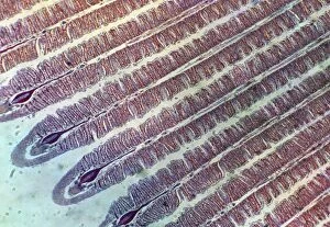
Microscopic Fish GillPM-1947 Fish Anatomy - Fish gill under microscope. Pat Morris Please note that prints are for personal display purposes only and may not be reproduced in anyway
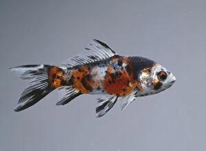
Shubumpkin BB 380 © John Daniels / ARDEA LONDONJD-13489 Calico SHUBUNKIN FISH / Goldfish John Daniels Please note that prints are for personal display purposes only and may not be reproduced in anyway
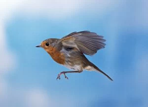
Robin - in flightBB-555 European ROBIN - side profile, in flight Erithacus rubecula Brian Bevan Please note that prints are for personal display purposes only and may not be reproduced in anyway
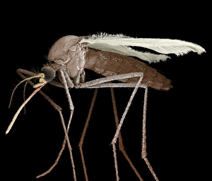
Scanning Electron Micrograph (SEM): Mosquito, Magnification x 35 (A4 size: 29. 7 cm width)LRDS-84 Mosquito Scanning Electron Micrograph (SEM) Culex pipiens Magnification x 35 (A4 size: 29.7 cm width) Coloured by hand to enhance natural features
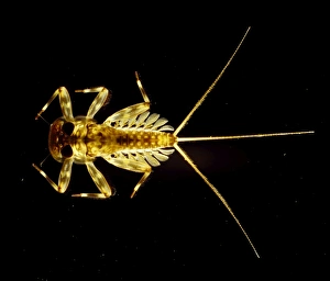
Light Micrograph : Flattened Mayfly Nymph, Magnification x 10 (A4 size: 29. 7 cm width)LRDS-80 Flattened Mayfly Nymph Light Micrograph Ecdyonurus venosus Magnification x 10 (A4 size: 29.7 cm width) Credit: David Spears (last refuge)
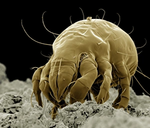
Scanning Electron Micrograph (SEM): Flour Mite, Magnification x 500 (A4 size: 29. 7 cm width)LRDS-58 Flour Mite Scanning Electron Micrograph (SEM) Acarus siro Magnification x 500 (A4 size: 29.7 cm width) Coloured by hand to enhance natural features
Scanning Electron Micrograph (SEM): Cat Flea, Magnification x250 (A4 size: 29. 7 cm width)LRDS-54 Cat Flea Scanning Electron Micrograph (SEM) Ctenocephalides felis Magnification x250 (A4 size: 29.7 cm width) Coloured by hand to enhance natural features
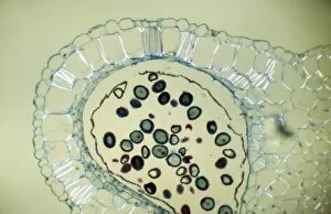
Light Micrograph (LM): Transverse section of Dehiscence Lilium Anthers with Pollen; Magnification x300 (on 10)LRDS-200 Light Micrograph (LM): Transverse section of Dehiscence Lilium Anthers with Pollen Magnification x300 (on 10.5 cm width print) David Spears (Last Refuge)
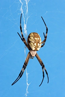
Golden Orb Weaver Spider - Size: abdomen only, 15 mm long; abdomen and headFG-eb-884 Golden Orb Weaver Spider Specimen from San Diego, California. Argiopes aurantia Size: abdomen only, 15 mm long; abdomen and head
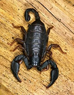
European Black Scorpion - This specimen found in a house in Reading, BerkshireSPH-3300 European Black Scorpion This specimen found in a house in Reading, UK - probably imported in holiday baggage Widespread in southern Europe Euscorpius italicus Introduced to UK via luggage
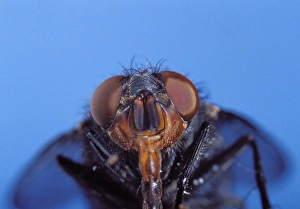
Blue Bottle fly - head with proboscis extendedPPG-1764 Bluebottle fly - head with proboscis extended Europe Calliphora vomitoria Pascal Goetgheluck Please note that prints are for personal display purposes only
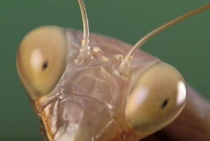
Praying Mantis - eyes EuropePPG-1654 Praying Mantis - eyes Europe Mantis religiosa Pascal Goetgheluck Please note that prints are for personal display purposes only and may not be reproduced in any way
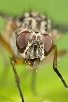
Muscidae Fly - Eyes and face Norfolk UKFEU-167 Muscidae Fly - Eyes and face Norfolk UK Phaonia sp. ? Geoff du Feu Please note that prints are for personal display purposes only and may not be reproduced in any way
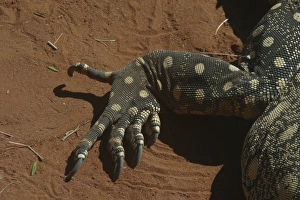
Perentie Goanna / Perenty Monitor Lizard Close up of foot Alice Springs, Nthn Territory, AustraliaDH-3153 Perentie Monitor Lizard / Goanna - Close up of foot Alice Springs, Nthn Territory, Australia Varanus giganteus Don Hadden Please note that prints are for personal display purposes only
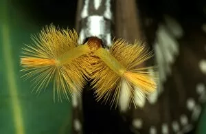
CLY02012AUS-234 Common crow butterfly - male with hair pencils everted to disperse sex pheromone Euploea core corinna New South Wales
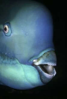
Bullethead Parrotfish - Note the many teeth that form the parrot like beakVT-8715 Bullethead Parrotfish - Note the many teeth that form the parrot like beak. These teeth are specially designed for munching coral Heron Island, Great Barrier Reef
SunflowerUSH-1065 SUNFLOWER - Seeds in ripened flower head Helianthus annuus Duncan Usher Please note that prints are for personal display purposes only and may not be reproduced in any way
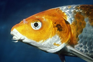
Koi CarpPM-7088 Koi Carp - Close up of head Cyprinus carpio Pat Morris Please note that prints are for personal display purposes only and may not be reproduced in any way
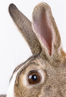
Rabbit - close-up of earsJD-10444 Rabbit - close-up of ears John Daniels Please note that prints are for personal display purposes only and may not be reproduced in any way
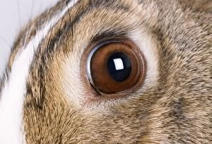
Rabbit - close-up of eyeJD-10442 Rabbit - close-up of eye John Daniels Please note that prints are for personal display purposes only and may not be reproduced in any way
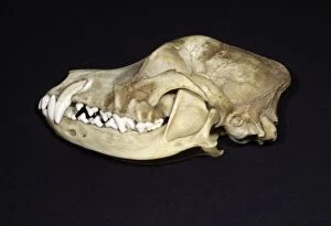
Dog's Skull Left side - Left side with bone over canine tooth removed to reveal the length of the rootHEN-18 Dog's Skull Left side - Left side with bone over canine tooth removed to reveal the length of the root. This explains why surgical removal of the canine tooth is difficult