mail_outline sales@mediastorehouse.com
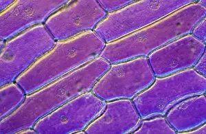
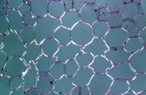
Light Micrograph (LM): Onion skin cells; Magnification x600 (on 10. 5 cm width print)LRDS-252 Light Micrograph (LM): Onion skin cells; Magnification x600 (on 10.5 cm width print) David Spears (Last Refuge) / ardea.com Last Refuge Please note that prints are for personal display
Light Micrograph (LM): Longitudinal section of stem of Crown of Thorns (Euphorbia splendens)LRDS-248 Light Micrograph (LM): Longitudinal section of stem of Crown of Thorns Magnification x1200 (on 10.5 cm width print) Euphorbia splendens David Spears (Last Refuge)
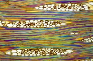
Light Micrograph (LM): Longitudinal section of Mahogani (Swietenia mahagoni); Magnification x600 (on 10)LRDS-246 Light Micrograph (LM): Longitudinal section of Mahogani Magnification x600 (on 10.5 cm width print) Swietenia mahagoni David Spears (Last Refuge)
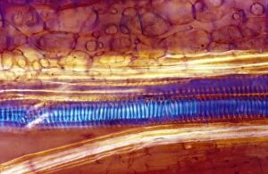
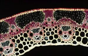
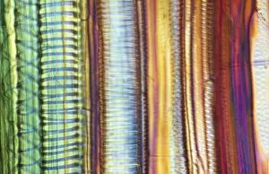
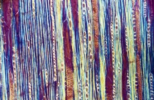
Light Micrograph (LM): Longitudinal section of Scots pine wood, (Pinus sylvestris); Magnification x480 (on 10)LRDS-245 Light Micrograph (LM): Longitudinal section of Scots pine wood, Magnification x480 (on 10.5 cm width print) Pinus sylvestris David Spears (Last Refuge)
Light Micrograph (LM): A transverse section of a straw of Wheat; Magnification x600 (on 10. 5 cm width print)LRDS-239 Light Micrograph (LM): A transverse section of a straw of Wheat Magnification x600 (on 10.5 cm width print) David Spears (Last Refuge)
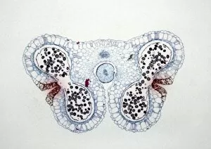
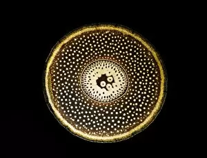
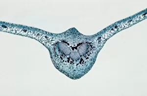
Light Micrograph (LM): Transverse section of Lilium Anthers with Mature Pollen; Magnification x15 (on 10)LRDS-234 Light Micrograph (LM): Transverse section of Lilium Anthers with Mature Pollen Magnification x15 (on 10.5 cm width print) David Spears (Last Refuge)
Light Micrograph (LM): A transverse section of a root of Conifer (Pandanus sp. ); Magnification x30 (on 10)LRDS-210 Light Micrograph (LM): A transverse section of a root of Conifer Magnification x30 (on 10.5 cm width print) Pandanus sp
Light Micrograph (LM): A transverse section of an aerial root of Orchid (Dendrobium sp. ); Magnification x30 (on 10)LRDS-209 Light Micrograph (LM): A transverse section of an aerial root of Orchid Magnification x30 (on 10.5 cm width print) Dendrobium sp
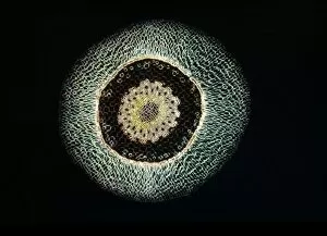
Light Micrograph (LM): A transverse section of a leaf of a Tulip (Tulipa sp)LRDS-202 Light Micrograph (LM): A transverse section of a leaf of a Tulip (Tulipa sp.) showing Stomata Magnification x600 (on 10.5 cm width print) Tulipa sp
Light Micrograph (LM): Longitudinal section of old sunflower stem(Helianthus sp)LRDS-199 Light Micrograph (LM): Longitudinal section of old sunflower stem(Helianthus sp.) showing spiral chloroplasts Magnification x1200 (on 10.5 cm width print)
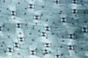
Light Micrograph (LM): A concentration of cells on the epidermis of a plant showing stomataLRDS-197 Light Micrograph (LM): A concentration of cells on the epidermis of a plant showing stomata David Spears (Last Refuge)
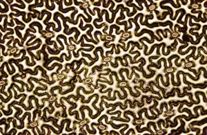
Light Micrograph (LM): Longitudinal section shows Scalariform vessels of Fern; Magnification x1200 (on 10)LRDS-196 Light Micrograph (LM): Longitudinal section shows Scalariform vessels of Fern Magnification x1200 (on 10.5 cm width print) David Spears (Last Refuge)
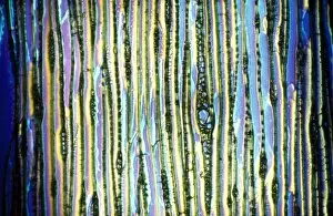
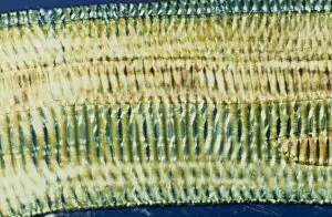
Light Micrograph (LM): A longitudinal section f a Ribes sp. stem; Magnification x600 (on 10. 5 cm width print)LRDS-194 Light Micrograph (LM): A longitudinal section of a Ribes sp. stem Magnification x600 (on 10.5 cm width print) Ribes sp
Light Micrograph (LM): transverse section of Prunus Leaf; Magnification x18 (on 10. 5 cm width print)LRDS-181 Light Micrograph (LM): transverse section of Prunus Leaf Magnification x18 (on 10.5 cm width print) Prunus sp David Spears (Last Refuge)
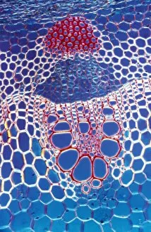
Light Micrograph (LM): A transverse section shows Vascular Bundle in Helianthus stem; Magnification x600 (on 10)LRDS-177 Light Micrograph (LM): A transverse section shows Vascular Bundle in Helianthus stem Magnification x600 (on 10.5 cm width print) Helianthus sp David Spears (Last Refuge)
Light Micrograph (LM): transvers section shows Parenchyma Ground Tissue in Buttercup stem; Magnification x600 (on 10)LRDS-176 Light Micrograph (LM): transvers section shows Parenchyma Ground Tissue in Buttercup stem Magnification x600 (on 10.5 cm width print) David Spears (Last Refuge)
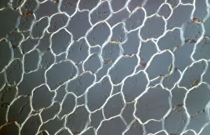
Light Micrograph (LM): tranverse section shows Parenchyma or packing tissue from Cucurbita Stem; Magnification x600LRDS-174 Light Micrograph (LM): tranverse section shows Parenchyma or packing tissue from Cucurbita Stem Magnification x600 (on 10.5 cm width print) David Spears (Last Refuge)
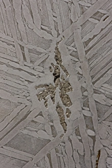
Seymchan Meteorite Cross-section - St. Petersburg Paleontological Lab - RussiaCAN-3456 Seymchan Meteorite Cross-section St. Petersburg Paleontological Lab - Russia John Cancalosi Please note that prints are for personal display purposes only
Gall Wasp - cross sectionPPG-285 Gall Wasp - cross section Biorrhiza pallida Pascal Goetgheluck Please note that prints are for personal display purposes only and may not be reproduced in any way
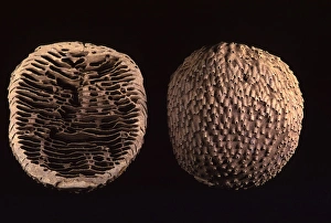
Termite - nest cut in halfPPG-1661 Termite - nest cut in half. Africa Apicotermes Pascal Goetgheluck Please note that prints are for personal display purposes only and may not be reproduced in any way
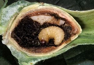
Hazelnut Weevil - larva in an hazelnut. EuropePPG-1614 Hazelnut Weevil - larva in an hazelnut Europe Curculio nucum Pascal Goetgheluck Please note that prints are for personal display purposes only and may not be reproduced in any way
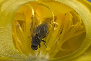
Globe flower - The Chiastocheta fly is inside the flower and will fertilize it. EuropePPG-1501 Globe flower - with Chiastocheta fly inside, cross-section Europe. Trollius europaeus The fly pollinates the host plant and then lays eggs on the developing fruit
Fossil - Mammoth Tooth - cross-section. Pleistocene South Carolina, USA E50T4155CAN-2181 Fossil - Mammoth Tooth - cross-section. Pleistocene South Carolina, USA Mammuthus meridionalis John Cancalosi Please note that prints are for personal display purposes only