mail_outline sales@mediastorehouse.com
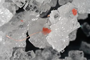
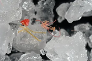
Picture No. 12479815Microplastics on table salt. Tiny fragments and Date:
Picture No. 12479814Microplastics on table salt. Tiny fragments and Date:
Picture No. 12479813Microplastics on table salt. Tiny fragments and Date:
Picture No. 12479812Microplastics on table salt. Tiny fragments and Date:
Picture No. 12479811Microplastics on table salt. Tiny fragments and Date:
Picture No. 12479810Microplastics on table salt. Tiny fragments and Date:
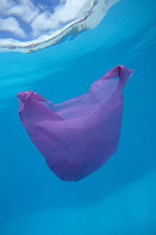
Picture No. 12479503Plastic bag driffting in the ocean. Plastic bags Date:
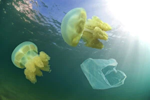
Picture No. 12479504Jellyfishes and plastic bag driffting. For us, Date:
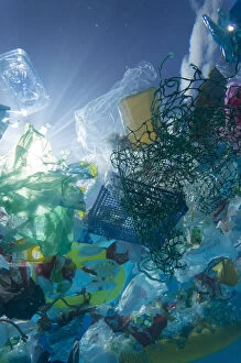
Picture No. 12479502Plastic garbage floating in the ocean. Unlike Date:
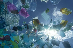
Picture No. 12479501Plastic garbage floating in the ocean. Unlike Date:
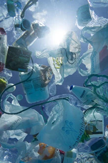
Picture No. 12479500Plastic garbage floating in the ocean. Unlike Date:
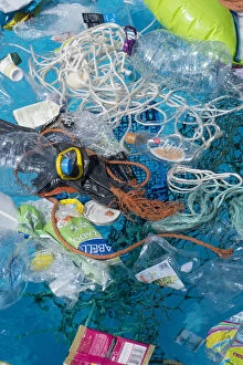
Picture No. 12479499Plastic garbage floating in the ocean. Unlike Date:
Picture No. 12479498Plastic garbage floating in the ocean. Unlike Date:
Picture No. 12479492Concept image to illustrate marine micoplastic Date:
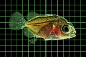
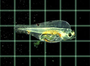
Picture No. 12479490Marine fish larvae eat microplastics. Small pieces Date:
Picture No. 12479491Marine fish larvae eat microplastics. Small pieces Date:
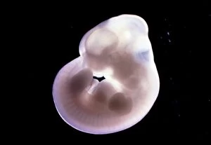
Picture No. 11675507Rat Embryo at 14.5 days old. Date:
Picture No. 11014634Common Frog - mid stage of tadpole development Scanning Electron Micrograph (SEM): Magnification x 85 (if print A4 size: 29.7 cm wide) - Gills are resorbed and jaws better developed
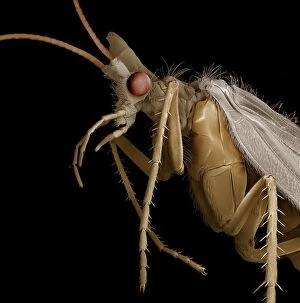
Picture No. 11014629Caddis Fly Scanning Electron Micrograph (SEM): Magnification x 20 (if print A4 size: 29.7 cm wide) - Caddis flies spend most of their lives as aquatic larvae in ponds and streams
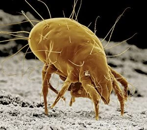
Picture No. 11014630Cheese Mite Scanning Electron Micrograph (SEM): Magnification x 350 (if print A4 size: 29.7 cm wide) (Tyrolichus casei) Date:
Picture No. 11014628Caenorhabditis elegans Scanning Electron Micrograph (SEM): Magnification x 1750 (if print A4 size: 29.7 cm wide) - This tiny free living nematode worm is extensively used in medical
Picture No. 11014624Escherishia Coli Bacteria / E. Coli - affected by antibiotic Scanning Electron Micrograph (SEM): Magnification x25, 000 (if print A4 size)
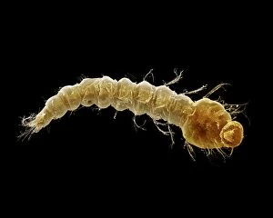
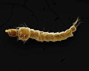
Picture No. 11014620Mosquito Larvae Scanning Electron Micrograph (SEM): Magnification x40 (if print A4 size: 29.7 cm wide) - The malarial mosquito larvae live in pools and puddles; almost any standing water will do
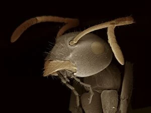
Picture No. 11014623Black Garden Ant Scanning Electron Micrograph (SEM): Magnification x120 (if print A4 size: 29.7 cm wide) (Lasius niger) Date:
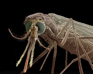
Picture No. 11014622Mosquito Scanning Electron Micrograph (SEM): Magnification x90 (if print A4 size: 29.7 cm wide) - Portrait of the malarial mosquito showing the antenae and palps
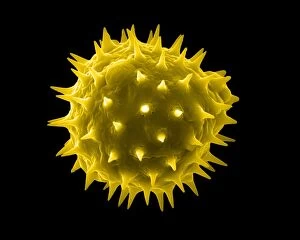
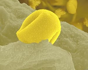
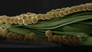
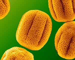
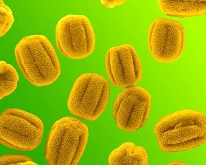
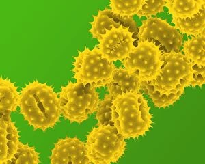
Picture No. 11014616Sunflower Pollen Scanning Electron Micrograph (SEM): Magnification x15, 000 (if print A4 size: 29.7 cm wide) - Sunflowers have huge inflorescences that shed large amounts of pollen that is carried
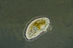
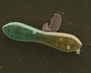
Picture No. 11014599Freshwater Ciliates common in pools and ponds consisting of a single cell and feed by ingesting other tiny organisms such as bacteria and other protozoa Date:
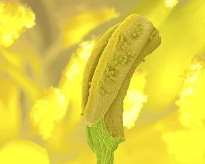
Picture No. 11014592Aconite Anther - with pollen Scanning Electron Micrograph (SEM): Magnification x 11150 (if print A4 size: 29.7 cm wide) - Aconites are attractive springtime garden flowers - They are insect
Picture No. 11014591Aconite Anther - with pollen Scanning Electron Micrograph (SEM): Magnification x 450 (if print A4 size: 29.7 cm wide) - Aconites are attractive springtime garden flowers - They are insect pollinated
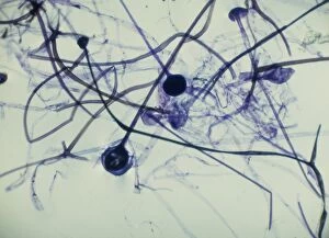
Picture No. 11675563Light Micrograph (LM): Rhizopus sporangia. Date:
Picture No. 11675503Rat Embryo 13.2 days after fertilisation. Date:
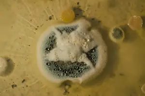
Picture No. 11014633Penicillin Colony This is a culture of the original strain of bacteria used by Flemming in the early research into the first antibiotics. Obliquely lit macro image from a time-lapse sequence. Date:
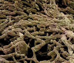
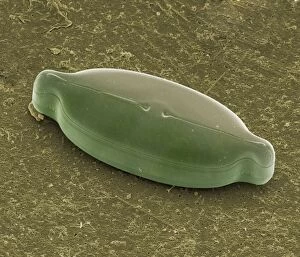
Picture No. 11014631Diatom Scanning Electron Micrograph (SEM): Magnification x5275 (if print A4 size: 29.7 cm wide) - The diatoms are single celled algae
Picture No. 11014632Diatom Scanning Electron Micrograph (SEM): Magnification x 4370 (if print A4 size: 29.7 cm wide) - The diatoms are single celled algae
Picture No. 11014627Spectacle Moth - eggs Scanning Electron Micrograph (SEM): Magnification x 35 (if print A4 size: 29.7 cm wide) (Abrostola tripartita) Date:
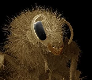
Picture No. 11014626Bumblebee Scanning Electron Micrograph (SEM): Magnification x30 (if print A4 size: 29.7 cm wide) (Bombus terrestris) Date:
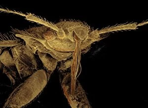
Picture No. 11014625Bedbug Scanning Electron Micrograph (SEM): Magnification x120 (if print A4 size: 29.7 cm wide) (Cimex lectularius) Date:
Picture No. 11014621Mosquito Larvae Scanning Electron Micrograph (SEM): Magnification x40 (if print A4 size: 29.7 cm wide) - The malarial mosquito larvae live in pools and puddles; almost any standing water will do
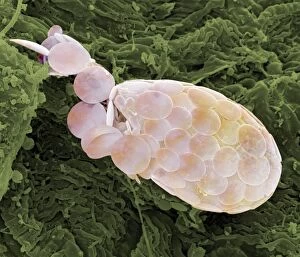
Picture No. 11014619Shelled Amoeba Scanning Electron Micrograph (SEM)): Magnification x5130 (if print A4 size: 29.7 cm wide) - Testate amoebae are found in damp soil, leaf litter and pools
Picture No. 11014614Quinidine Liquified Quinidine is a chemical precurser to the antimalarial drug quinine - The image was produced using a polarising microscope and post processed in Adobe Photoshop Date:
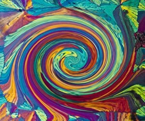
Picture No. 11014613Quinidine Swirl Quinidine is a chemical precurser to the antimalarial drug quinine - The image was produced using a polarising microscope and post processed in Adobe Photoshop Date:
Picture No. 11014611Primrose Pollen Scanning Electron Micrograph (SEM): Magnification 2 x28, 000 (if print A4 size: 29.7 cm wide) - Primroses appear in early spring and are usually yellow
Picture No. 11014610Primrose Pollen Scanning Electron Micrograph (SEM): Magnification x12, 000 (if print A4 size: 29.7 cm wide) - Primroses appear in early spring and are usually yellow. They are insect pollinated Date:
Picture No. 11014609Hazel Pollen Scanning Electron Micrograph (SEM): Magnification x12, 000 b1 (if print A4 size: 29.7 cm wide) - Hazel pollen is very small and is carried by the wind to stigma of other trees
Picture No. 11014608Grass Pollen Scanning Electron Micrograph (SEM): Magnification 2 x4560 (if print A4 size: 29.7 cm wide) - Gypsophyla is a small pink or white flower; insect pollinated Date:
Picture No. 11014605Grass Pollen Scanning Electron Micrograph (SEM): Magnification 2 x9410 (if print A4 size: 29.7 cm wide) - Grass pollen is wind pollinated
Picture No. 11014602Daffodil Pollen Scanning Electron Micrograph (SEM): Magnification x16900 (if print A4 size: 29.7 cm wide) - Daffodils are popular and common springtime flowers. Insect pollinated Date:
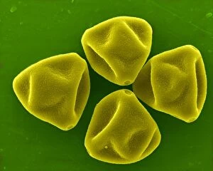
Picture No. 11014603Daisy Pollen Scanning Electron Micrograph (SEM): Magnification 2 x9k (if print A4 size: 29.7 cm wide) - Daisies are small insect pollinated flowers; common in lawns Date: