mail_outline sales@mediastorehouse.com
Picture No. 11014617Veronica Pollen Scanning Electron Micrograph (SEM): Magnification x8220 (if print A4 size: 29.7 cm wide) - These small pollen grains are sticky and are carried away by pollinating insects Date:
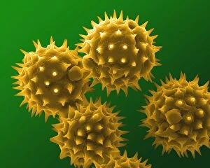
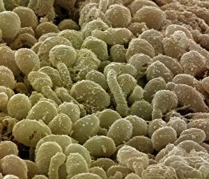
Picture No. 11014615Sunflower Pollen Scanning Electron Micrograph (SEM): Magnification x10, 000 (if print A4 size: 29.7 cm wide) - The large flowers are insect pollinated
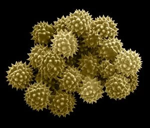
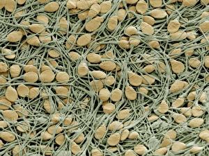
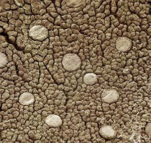
Picture No. 11014607Groundsel Pollen Scanning Electron Micrograph (SEM): Magnification 2 x6940 (if print A4 size: 29.7 cm wide) - Groundsel is a common weed - It is insect pollinated; provides food for many insects
Picture No. 11014606Groundsel Pollen Scanning Electron Micrograph (SEM): Magnification 2 x6940 (if print A4 size: 29.7 cm wide) - Groundsel is a common weed - It is insect pollinated; provides food for many insects
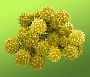
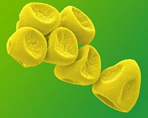
Picture No. 11014604Gorse Pollen Scanning Electron Micrograph (SEM): Magnification 1 x10, 000 (if print A4 size: 29.7 cm wide) - Gorse is a bush, with brigh yellow flowers in spring and early summer
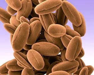
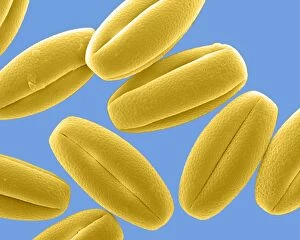
Picture No. 11014601Daffodil Pollen Scanning Electron Micrograph (SEM): Magnification x8930 (if print A4 size: 29.7 cm wide) - Daffodils are popular and common springtime flowers. Insect pollinated Date:
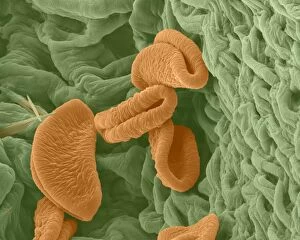
Picture No. 11014593Peruvian Lily / Alstroemeria Pollen on Anther Scanning Electron Micrograph (SEM): Magnification x6520 (if print A4 size: 29.7 cm wide)
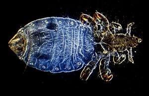
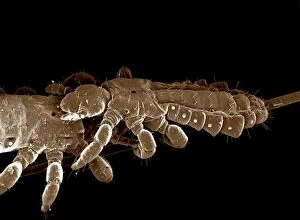
Picture No. 10873636Human Head Louse (Pediculus humanus capitis) Date:
Picture No. 10874130Light Micrograph (LM): Hookworm (Ancylostoma duodenale) Date:
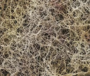
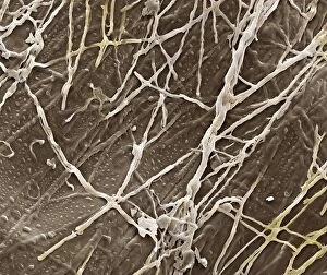
Picture No. 10873581Athleteճ Foot Fungus (Tinea pedis) Date:
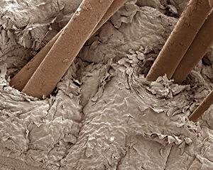
Picture No. 10873643Human scalp Date:
Picture No. 10873686Human sperm cells Date:
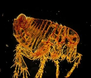
Picture No. 10873644Human Flea - male (Pulex irritans) Date:
Picture No. 10873600Human Crab Louse (Phthirus pubis) Date:
Picture No. 10873580Athleteճ Foot Fungu (Tinea pedis) Date:
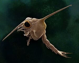
Picture No. 10873601Crab larvae (Zoea larvae) Date:
Picture No. 10873638Human Head Louse (Pediculus humanus capitis) Date:
Picture No. 10873634Cochlea of Guinea Pig Date:
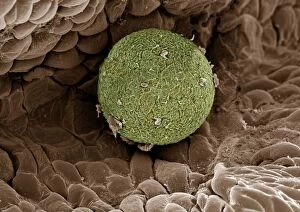
Picture No. 10873615Human Ovum in Fallopian Tube : This freshly fertilized egg passes down the Fallopian tube on its way to implantation in the wall of the uterus ( ) Date:
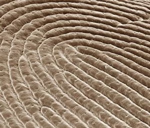
Picture No. 10873617Human finger tip print Date:
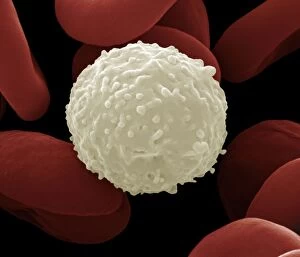
Picture No. 10873675Human White and Red Blood Cells Date:
Picture No. 10873616Human Ovum in Fallopian Tube : This freshly fertilized egg passes down the Fallopian tube on its way to implantation in the wall of the uterus Date:
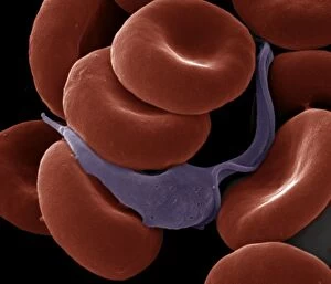
Picture No. 10873703Sleeping Sickness Parasite in red blood cells (Trypanosoma sp.) Date:
Picture No. 10873596Human Cochlea Date:
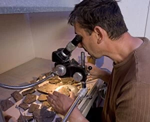
Picture No. 10891096Paleontologist / Palaeontologist Eric Depre using microscope in workshop examining fossils Date:
Picture No. 10877011Scanning Electron Micrograph (SEM): Frog tongue - taste receptors papillae Date:
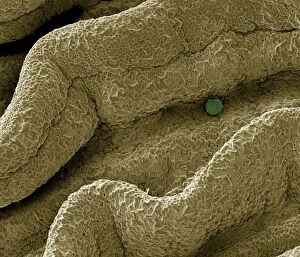
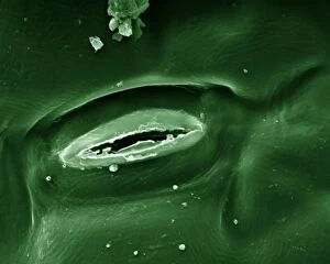
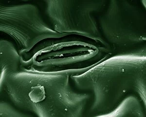
Picture No. 10877020Scanning Electron micrograph (SEM): Open Stoma of a plant leaf; Date:
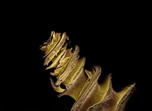
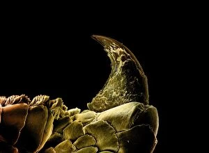
Picture No. 10877013Scanning Electron Micrograph (SEM); Gecko Claw: Date:
Picture No. 10877021Scanning Electron micrograph (SEM): Open Stoma of a plant leaf; Date:
Picture No. 10877014Scanning Electron Micrograph (SEM): Gecko foot hairs Date:
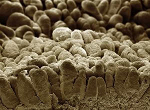
Picture No. 10877012Scanning Electron Micrograph (SEM): human intestine showing villi Date:
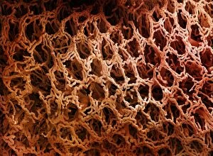
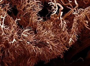
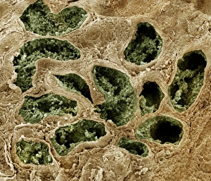
Picture No. 10877008Scanning Electron Micrograph (SEM) of a resin cast of blood vessels in the intestines; Date:
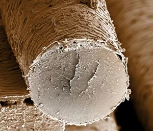
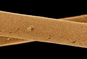
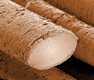
Picture No. 10877005Scanning Electron Micrograph (SEM): Human hair - Asian Date:
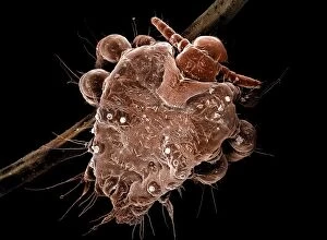
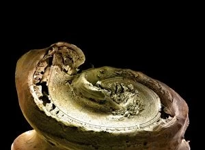
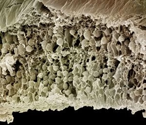
Picture No. 10877009Scanning Electron Micrograph (SEM): a corrosion cast of a gut tumour - Liquid plastic is injected into the blood vessels; it solidifies
Picture No. 10877007Scanning Electron Micrograph (SEM): Human Hairs Date:
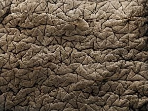
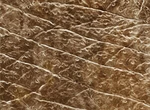
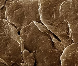
Picture No. 10876998Scanning Electron Micrograph (SEM): Human Skin Date:
Picture No. 10877000Scanning Electron Micrograph (SEM): Human Skin Date:
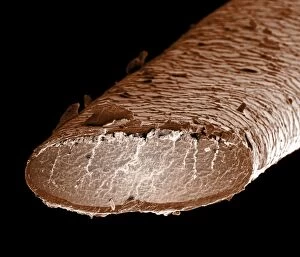
Picture No. 10877004Scanning Electron Micrograph (SEM): Human hair - European Date:
Picture No. 10877006Scanning Electron Micrograph (SEM): Human pubic hair Date:
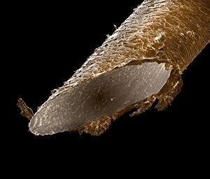
Picture No. 10877003Scanning Electron Micrograph (SEM): Human hair - African Date:
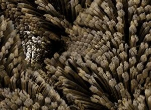
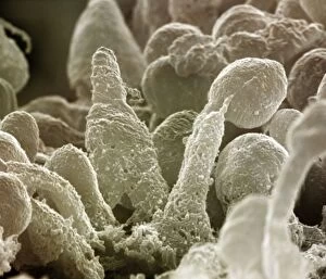
Picture No. 10876996Scanning Electron Micrograph (SEM): Human Retina showing photoreceptors rod and cone cells Date:
Picture No. 10877001Scanning Electron Micrograph (SEM): Human Skin Date:
Picture No. 10876999Scanning Electron Micrograph (SEM): Human Skin Date:
Picture No. 10876991Scanning Electron Micrograph (SEM): Human testis tubes - Sperm cells forming in seminal tubes of testis Date:
Picture No. 10876993Scanning Electron Micrograph (SEM): Human Retina showing photoreceptors rod and cone cells Date:
Picture No. 10876995Scanning Electron Micrograph (SEM): Human Retina showing photoreceptors rod and cone cells Date: