mail_outline sales@mediastorehouse.com
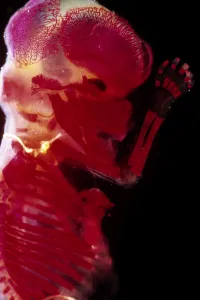
Picture No. 10875775Light Micrograph of Human Foetus 12 weeks - red dye to show bones Date:
Picture No. 10875778Light Micrograph of Human Embryo With Placenta Date:
Picture No. 10875776Light Micrograph of Human Foetus 12 weeks - red dye to show bones Date:
Picture No. 10875777Light Micrograph of Human Foetus 10-11 weeks after fertilisation Date:
Picture No. 10875774Light Micrograph of Human Foetus 12 weeks - red dye to show bones; Date:
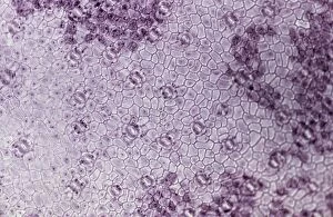
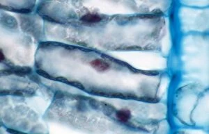
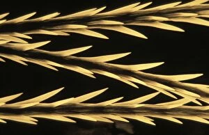
Picture No. 10874465Light Micrograph (LM): A transverse section of a lower Epidermis of a Box leaf (Buxus sp.) showing Stomata (Buxus sp.) Date:
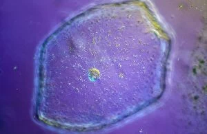
Picture No. 10874442Light Micrograph (LM): Tomato seed; Magnification x600 (on 10.5 cm width print) Date:
Picture No. 10874441Light Micrograph (LM): Tomato seed; Magnification x600 (on 10.5 cm width print) Date:
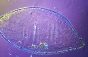
Picture No. 10874543Scanning Electron Micrograph (SEM): Tapeworm from a teleost fish Date:
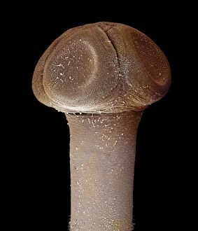
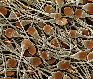
Picture No. 10874544Scanning Electron Micrograph (SEM): human sperm Date:
Picture No. 10874514Light Micrograph (LM): A light micrograph of the nuclei of plant cells Date:
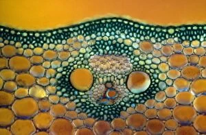
Picture No. 10874466Light Micrograph (LM): A transverse section of a Maize stem (Zea sp.) (Zea sp) Date:
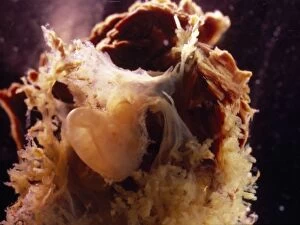
Picture No. 10895903Tarantula - Urticants hair under SEM (Scanning Electron Microscope) (Lasiodora parahybana) Date:
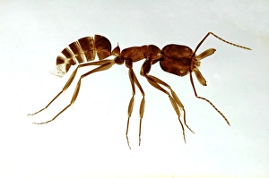
Ant, made as a cleared and flattened microscope preparationPM-10629 Ant, made as a cleared and flattened microscope preparation. Pat Morris Please note that prints are for personal display purposes only and may not be reproduced in any way