mail_outline sales@mediastorehouse.com
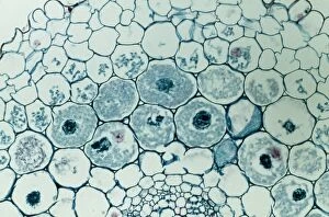
Light Micrograph (LM): Filaments of fungi Endotrophic Mycorrhiza live within cells of a root; Magnification x600LRDS-254 Light Micrograph (LM): Filaments of fungi Endotrophic Mycorrhiza live within cells of a root Magnification x600 (on 10.5 cm width print) David Spears (Last Refuge)
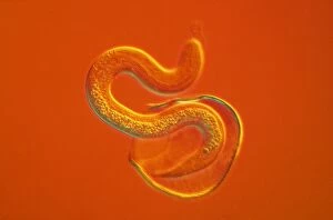
Light Micrograph - a roundworm hatching from its egg against an orange background. CHI0515LRDS-162 Light Micrograph - a roundworm hatching from its egg against an orange background. David Spears (Last Refuge) / ardea.com Last Refuge contact details: prints@ardea.com tel
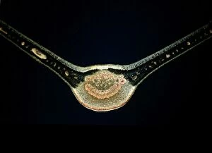
Bryozoa - moss animals. Microscopic x3 magnificationJC-249 Bryozoa - moss animals Microscopic x3 magnification Lophopus crytsallinus John Clegg Please note that prints are for personal display purposes only and may not be reproduced in any way
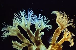
Bryozoa - microscopic, x10 magnificationJC-592 Bryozoa - microscopic, x10 magnification Bugula flabellaria John Clegg Please note that prints are for personal display purposes only and may not be reproduced in any way
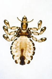
Pig Louse - x5 magnificationJC-253 Pig Louse - x5 magnification John Clegg Please note that prints are for personal display purposes only and may not be reproduced in any way
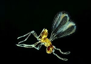
Fairy Fly - x 20 magnification family: ChalcididaeJC-238 Fairy Fly - x 20 magnification Eustochus sp. family: Chalcididae John Clegg Please note that prints are for personal display purposes only and may not be reproduced in any way
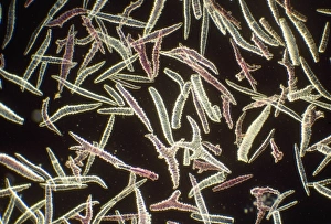
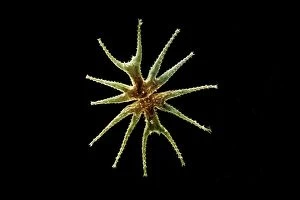
Sponge Spicules - microscopic, x4 magnificationJC-673 Sponge Spicules - microscopic, x4 magnification Gorgonia sp. John Clegg Please note that prints are for personal display purposes only and may not be reproduced in any way
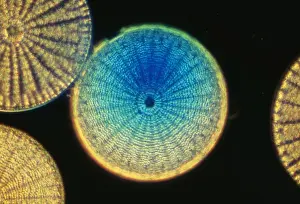
Diatoms SPH 523 From marine plankton sample. Santa Anna, California USA. © Steve Hopkin / ARDEA LONDONSPH-523 DIATOMS - From marine plankton sample. A type of phytoplankton Santa Anna, California USA Steve Hopkin Microscopic Please note that prints are for personal display purposes only
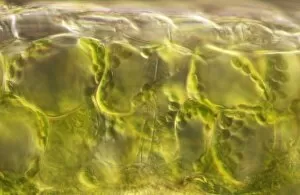
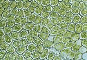
Light Micrograph (LM): plant cell chloroplasts - the site where photosynthesis takes placeLRDS-192 Light Micrograph (LM): plant cell chloroplasts - the site where photosynthesis takes place David Spears (Last Refuge)
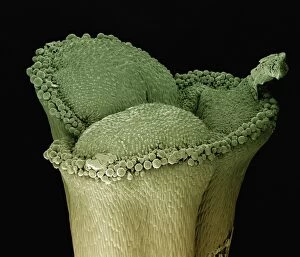
Scanning Electron Micrograph (SEM): stigma of a lily, Magnification x 100 (A4 size: 29. 7 cm width)LRDS-125 stigma of a lily Scanning Electron Micrograph (SEM) Lilium sp. Magnification x 100 (A4 size: 29.7 cm width) Coloured by hand to enhance natural features
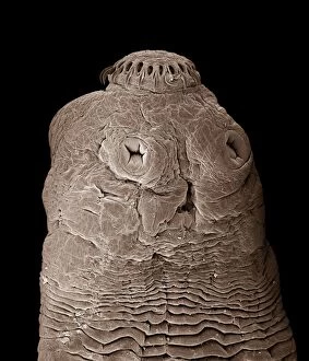
Tapeworm CHI0860LRDS-154 Tapeworm Taenia solium David Spears (Last Refuge) / ardea.com Last Refuge Please note that prints are for personal display purposes only and may not be reproduced in any way
Scanning Electron Micrograph (SEM): Calanus sp. ; Magnification x650 (A4 size: 29. 7 cm width)LRDS-302 Scanning Electron Micrograph (SEM): Calanus sp Magnification x650 (A4 size: 29.7 cm width) David Spears (Last Refuge)
Freshwater DesmidLRDS-303 Scanning Electron Micrograph (SEM): Freshwater Desmid Magnification x260 (when printed 10.5 cm Micrasterias lux David Spears (Last Refuge)
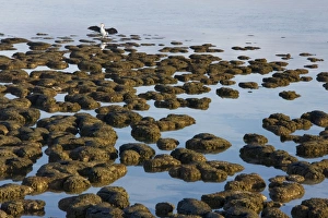
Stromatolites at Hamelin Pool, Shark Bay, western Australia. These are living representatives of the oldest livingROG-13733 Stromatolites at Hamelin Pool, Shark Bay, western Australia These are living representatives of the oldest living organisms, over 3.5 billion years old as fossils
Scanning Electron Micrograph (SEM): Groundsel, Magnification x 3, 000 (A4 size: 29. 7 cm width)LRDS-98 Groundsel Scanning Electron Micrograph (SEM) Senecio vulgaris Magnification x 3, 000 (A4 size: 29.7 cm width) Coloured by hand to enhance natural features
Light Micrograph (LM): Cells from a hair on the stamen of the common spiderwort (Tradescantia)LRDS-171 Light Micrograph (LM): Cells from a hair on the stamen of the common spiderwort (Tradescantia) Tradescantia sp David Spears (Last Refuge)
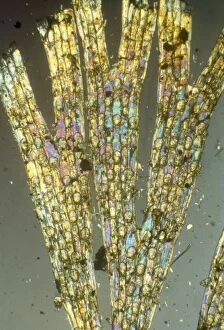
Light Micrograph (LM): A transverse section of a Fig leaf); Magnification x15 (on 10. 5 cm width print)LRDS-187 Light Micrograph (LM): A transverse section of a Fig leaf) Magnification x15 (on 10.5 cm width print) David Spears (Last Refuge)
Light Micrograph (LM): The cellular struture of a liverwort plant (Hepatica); Magnification x1200 (on 10)LRDS-190 Light Micrograph (LM): The cellular struture of a liverwort plant (Hepatica) Magnification x1200 (on 10.5 cm width print) Hepatica sp David Spears (Last Refuge)
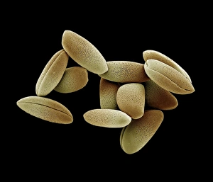
Scanning Electron Micrograph (SEM): Lily Pollen, Magnification x 1, 200 (A4 size: 29. 7 cm width) LRDS-99 Lily Pollen Scanning Electron Micrograph (SEM) Lilium sp. Magnification x 1, 200 (A4 size: 29.7 cm width) Coloured by hand to enhance natural features
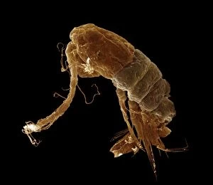
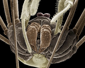
Scanning Electron Micrograph (SEM): Harvestman, Magnification x 70 (A4 size: 29. 7 cm width)LRDS-66 Harvestman Scanning Electron Micrograph (SEM) Phalangium opilio Magnification x 70 (A4 size: 29.7 cm width) Coloured by hand to enhance natural features
Light Micrograph (LM): A single cell from a hair on the stamen of the common spiderwort (Tradescantia)LRDS-170 Light Micrograph (LM): A single cell from a hair on the stamen of the common spiderwort (Tradescantia) Tradescantia sp David Spears (Last Refuge)