mail_outline sales@mediastorehouse.com
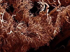
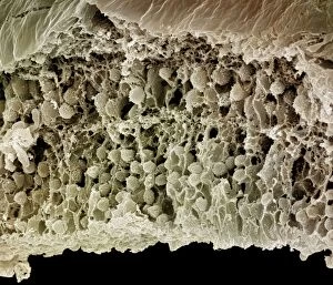
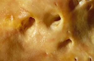
Picture No. 10877009Scanning Electron Micrograph (SEM): a corrosion cast of a gut tumour - Liquid plastic is injected into the blood vessels; it solidifies
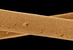
Picture No. 10877007Scanning Electron Micrograph (SEM): Human Hairs Date:
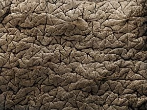
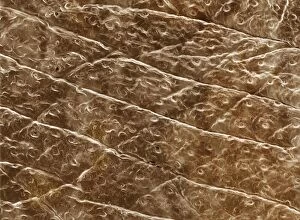
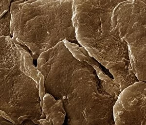
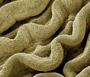
Picture No. 10876998Scanning Electron Micrograph (SEM): Human Skin Date:
Picture No. 10877000Scanning Electron Micrograph (SEM): Human Skin Date:
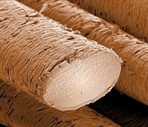
Picture No. 10877004Scanning Electron Micrograph (SEM): Human hair - European Date:
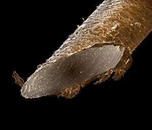
Picture No. 10877006Scanning Electron Micrograph (SEM): Human pubic hair Date:
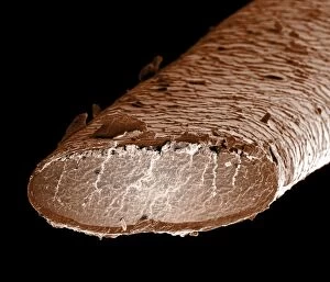
Picture No. 10877003Scanning Electron Micrograph (SEM): Human hair - African Date:
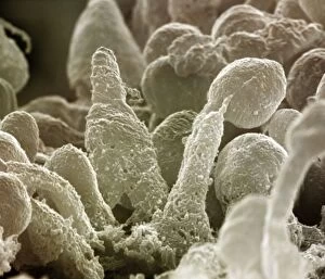
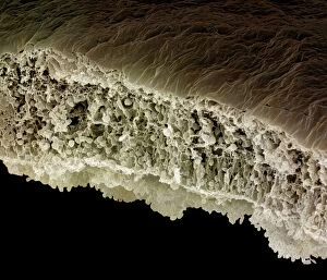
Picture No. 10876996Scanning Electron Micrograph (SEM): Human Retina showing photoreceptors rod and cone cells Date:
Picture No. 10877001Scanning Electron Micrograph (SEM): Human Skin Date:
Picture No. 10876999Scanning Electron Micrograph (SEM): Human Skin Date:
Picture No. 10876991Scanning Electron Micrograph (SEM): Human testis tubes - Sperm cells forming in seminal tubes of testis Date:
Picture No. 10876993Scanning Electron Micrograph (SEM): Human Retina showing photoreceptors rod and cone cells Date:
Picture No. 10876995Scanning Electron Micrograph (SEM): Human Retina showing photoreceptors rod and cone cells Date:
Picture No. 10876989Scanning Electron Micrograph (SEM): Human Fallopian tube Date:
Picture No. 10876994Scanning Electron Micrograph (SEM): Human Retina showing photoreceptors rod and cone cells Date:
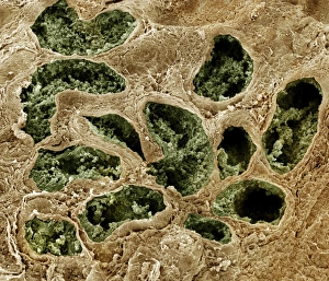
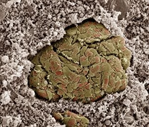
Picture No. 10876992Scanning electron micrograph (SEM) of Kidney glomerulus Date:
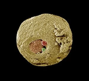
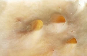
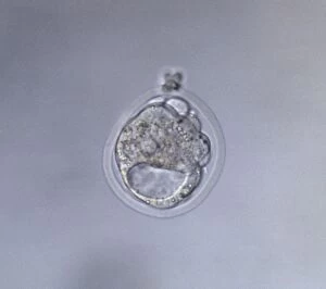
Picture No. 10876988Scanning Electron Micrograph (SEM): Early human embryo, one cell removed for genetic analysis Date:
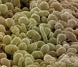
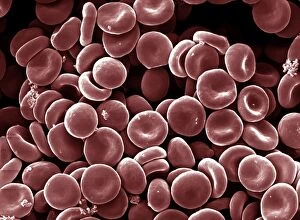
Picture No. 10876986Scanning Electron Micrograph (SEM): Human Red Blood Cells Date:
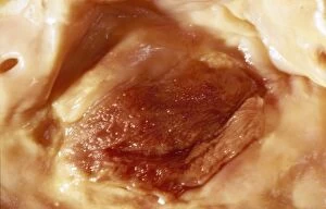
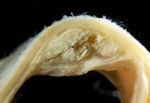
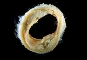
Picture No. 10875840Light Micrograph of Atherosclerotic Plaque and Obstructed Lumen of Human Aorta Date:
Picture No. 10875844Light Micrograph of Atherosclerotic Plaque and Obstructed Lumen of Human Aorta Date:
Picture No. 10875843Light Micrograph of Atherosclerotic Plaque and Obstructed Lumen of Human Aorta Date:
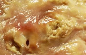
Picture No. 10875841Light Micrograph of Atherosclerotic Aorta Vessel Date:
Picture No. 10875839Light Micrograph of Atherosclerotic Plaque and Obstructed Lumen of Human Aorta Date:
Picture No. 10875842Light Micrograph of Atherosclerotic Plaque and Obstructed Lumen of Human Aorta Date:
Picture No. 10875838Light Micrograph of Atherosclerotic Plaque and Obstructed Lumen of Human Aorta Date:
Picture No. 10875837Light Micrograph of Atherosclerotic Plaque and Obstructed Lumen of Human Aorta Date:
Picture No. 10875836Light Micrograph of Atherosclerotic Plaque and Obstructed Lumen of Human Aorta Date:
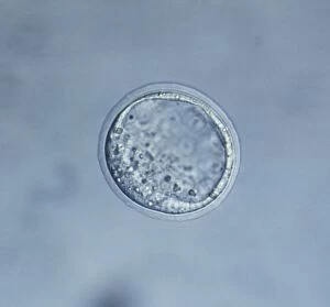
Picture No. 10875812Light Micrograph of Cavitated Mouse Blastocyst Date:
Picture No. 10875811Light Micrograph of Mouse Blastocyst Date:
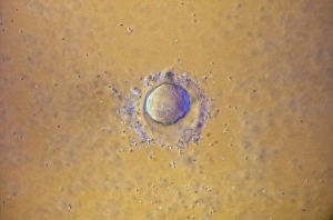
Picture No. 10875805Light Micrograph of Mouse Ovum being fertilized Date:
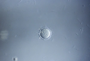
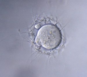
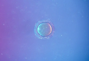
Picture No. 10875802Light Micrograph of human Ovum surrounded by Sperm - being fertilized Date:
Picture No. 10875806Light Micrograph of Mouse Ovum being fertilized Date:
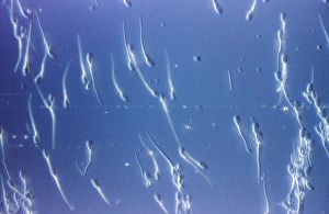
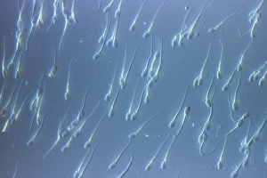
Picture No. 10875800Light Micrograph of Human Sperm; Date:
Picture No. 10875803Light Micrograph of human Ovum surrounded by Sperm - being fertilized Date:
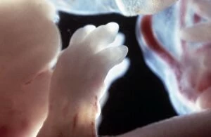
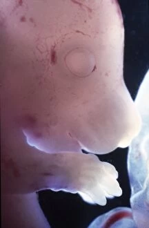
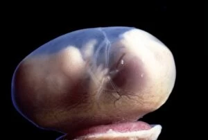
Picture No. 10875797Light Micrograph of Rat Embryo - The paws of a 17.5 day old Date:
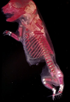
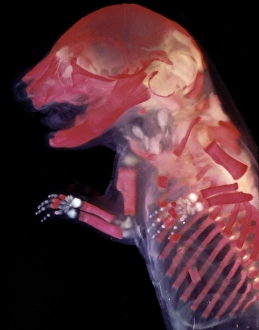
Picture No. 10875799Light Micrograph of Mouse Embryo - red dye to show bones Date:
Picture No. 10875801Light Micrograph of Human Sperm Date:
Picture No. 10875798Light Micrograph of Mouse Embryo - red dye to show bones Date:
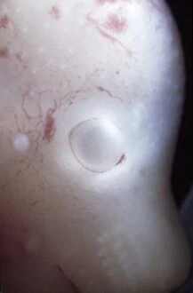
Picture No. 10875794Light Micrograph of Rat Embryo without its yolk sac, at 17.5 days Date:
Picture No. 10875795Light Micrograph of Rat Embryo without its yolk sac, at 17.5 days Date:
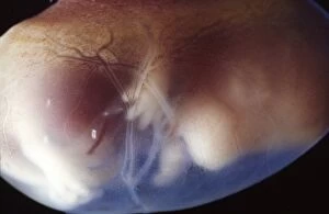
Picture No. 10875796Light Micrograph of Rat Embryo 17.5 days into its gestation period, in sac with placenta Date:
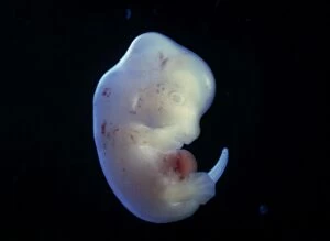
Picture No. 10875792Light Micrograph of Rat Embryo - 15.5 days into its gestation period - in sac with placenta Date:
Picture No. 10875790Light Micrograph of Rat Embryo - at 15.5 days, no sac, no placenta Date: