mail_outline sales@mediastorehouse.com
Picture No. 11014628Caenorhabditis elegans Scanning Electron Micrograph (SEM): Magnification x 1750 (if print A4 size: 29.7 cm wide) - This tiny free living nematode worm is extensively used in medical
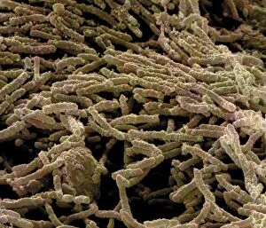
Picture No. 11014624Escherishia Coli Bacteria / E. Coli - affected by antibiotic Scanning Electron Micrograph (SEM): Magnification x25, 000 (if print A4 size)
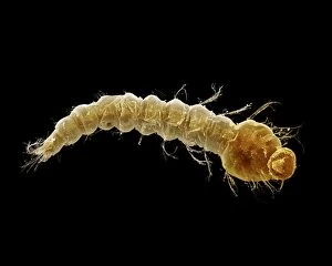
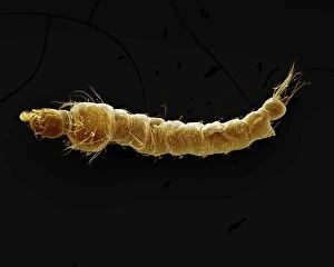
Picture No. 11014620Mosquito Larvae Scanning Electron Micrograph (SEM): Magnification x40 (if print A4 size: 29.7 cm wide) - The malarial mosquito larvae live in pools and puddles; almost any standing water will do
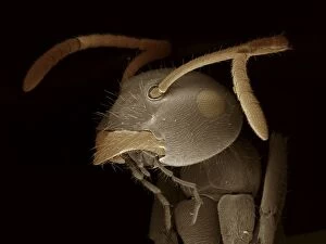
Picture No. 11014623Black Garden Ant Scanning Electron Micrograph (SEM): Magnification x120 (if print A4 size: 29.7 cm wide) (Lasius niger) Date:
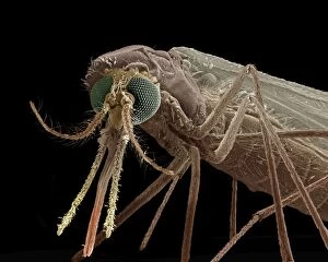
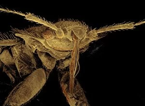
Picture No. 11014622Mosquito Scanning Electron Micrograph (SEM): Magnification x90 (if print A4 size: 29.7 cm wide) - Portrait of the malarial mosquito showing the antenae and palps
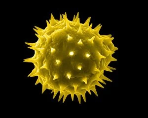
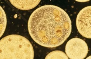
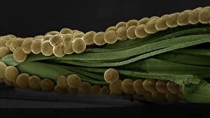
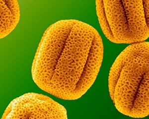
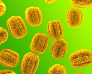
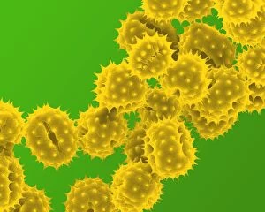
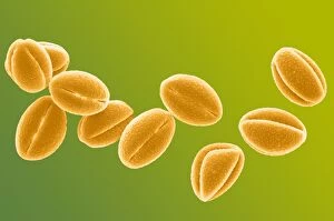
Picture No. 11014616Sunflower Pollen Scanning Electron Micrograph (SEM): Magnification x15, 000 (if print A4 size: 29.7 cm wide) - Sunflowers have huge inflorescences that shed large amounts of pollen that is carried
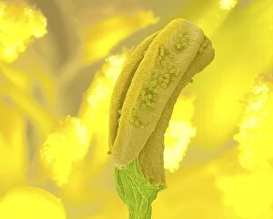
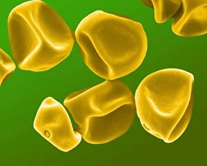
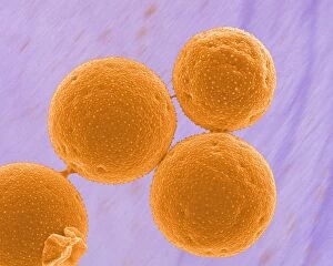
Picture No. 11014592Aconite Anther - with pollen Scanning Electron Micrograph (SEM): Magnification x 11150 (if print A4 size: 29.7 cm wide) - Aconites are attractive springtime garden flowers - They are insect
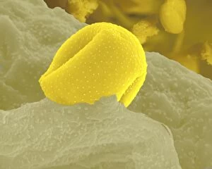
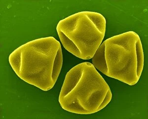
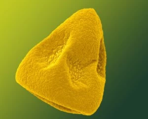
Picture No. 11014591Aconite Anther - with pollen Scanning Electron Micrograph (SEM): Magnification x 450 (if print A4 size: 29.7 cm wide) - Aconites are attractive springtime garden flowers - They are insect pollinated
Picture No. 10873637Human Head Louse (Pediculus humanus capitis) Date:
Colonial Green Algae SPH 2999 Volvox sp. © Steve Hopkin / ardea. comSPH-2999 Colonial Green ALGAE Volvox sp. Steve Hopkin Microscopic contact details: prints@ardea.com tel: +44 (0) 20 8318 1401
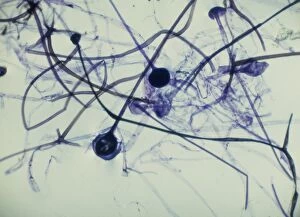
Picture No. 11675563Light Micrograph (LM): Rhizopus sporangia. Date:
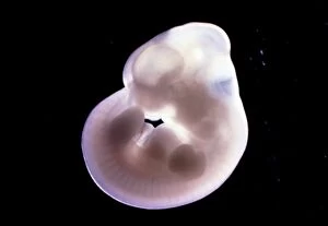
Picture No. 11675503Rat Embryo 13.2 days after fertilisation. Date:
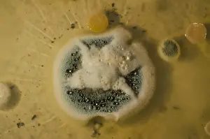
Picture No. 11014633Penicillin Colony This is a culture of the original strain of bacteria used by Flemming in the early research into the first antibiotics. Obliquely lit macro image from a time-lapse sequence. Date:
Picture No. 11014631Diatom Scanning Electron Micrograph (SEM): Magnification x5275 (if print A4 size: 29.7 cm wide) - The diatoms are single celled algae
Picture No. 11014632Diatom Scanning Electron Micrograph (SEM): Magnification x 4370 (if print A4 size: 29.7 cm wide) - The diatoms are single celled algae
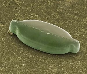
Picture No. 11014627Spectacle Moth - eggs Scanning Electron Micrograph (SEM): Magnification x 35 (if print A4 size: 29.7 cm wide) (Abrostola tripartita) Date:
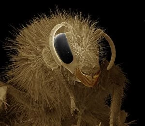
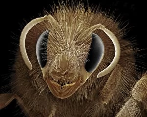
Picture No. 11014626Bumblebee Scanning Electron Micrograph (SEM): Magnification x30 (if print A4 size: 29.7 cm wide) (Bombus terrestris) Date:
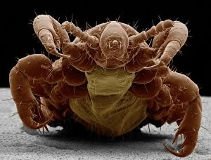
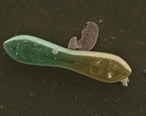
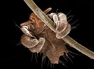
Picture No. 11014625Bedbug Scanning Electron Micrograph (SEM): Magnification x120 (if print A4 size: 29.7 cm wide) (Cimex lectularius) Date:
Picture No. 11014621Mosquito Larvae Scanning Electron Micrograph (SEM): Magnification x40 (if print A4 size: 29.7 cm wide) - The malarial mosquito larvae live in pools and puddles; almost any standing water will do
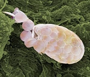
Picture No. 11014619Shelled Amoeba Scanning Electron Micrograph (SEM)): Magnification x5130 (if print A4 size: 29.7 cm wide) - Testate amoebae are found in damp soil, leaf litter and pools
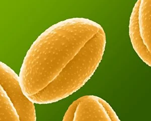
Picture No. 11014611Primrose Pollen Scanning Electron Micrograph (SEM): Magnification 2 x28, 000 (if print A4 size: 29.7 cm wide) - Primroses appear in early spring and are usually yellow
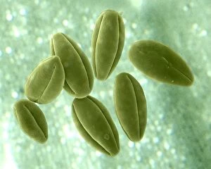
Picture No. 11014610Primrose Pollen Scanning Electron Micrograph (SEM): Magnification x12, 000 (if print A4 size: 29.7 cm wide) - Primroses appear in early spring and are usually yellow. They are insect pollinated Date:
Picture No. 11014609Hazel Pollen Scanning Electron Micrograph (SEM): Magnification x12, 000 b1 (if print A4 size: 29.7 cm wide) - Hazel pollen is very small and is carried by the wind to stigma of other trees
Picture No. 11014608Grass Pollen Scanning Electron Micrograph (SEM): Magnification 2 x4560 (if print A4 size: 29.7 cm wide) - Gypsophyla is a small pink or white flower; insect pollinated Date:
Picture No. 11014605Grass Pollen Scanning Electron Micrograph (SEM): Magnification 2 x9410 (if print A4 size: 29.7 cm wide) - Grass pollen is wind pollinated
Picture No. 11014602Daffodil Pollen Scanning Electron Micrograph (SEM): Magnification x16900 (if print A4 size: 29.7 cm wide) - Daffodils are popular and common springtime flowers. Insect pollinated Date:
Picture No. 11014603Daisy Pollen Scanning Electron Micrograph (SEM): Magnification 2 x9k (if print A4 size: 29.7 cm wide) - Daisies are small insect pollinated flowers; common in lawns Date:
Picture No. 11014600Crocus Pollen Scanning Electron Micrograph (SEM): Magnification x4000 (if print A4 size: 29.7 cm wide) - Crocus is a common spring flower, insect pollinated Date:
Picture No. 11014598Celandine Pollen Scanning Electron Micrograph (SEM): Magnification x17, 600 (if print A4 size: 29.7 cm wide) - is a yellow springtime flower. Insect pollinated, it is toxic to humans Date:
Picture No. 11014597Celandine Pollen Scanning Electron Micrograph (SEM): Magnification x4900 (if print A4 size: 29.7 cm wide) - is a yellow springtime flower. Insect pollinated, it is toxic to humans. Date:
Picture No. 11014595Bumblebee Scanning Electron Micrograph (SEM): Magnification x40 (if print A4 size: 29.7 cm wide) (Bombus terrestris) Date:
Picture No. 10873599Human Crab Louse (Phthirus pubis) Date:
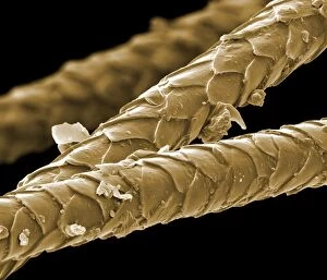
Picture No. 10877002Scanning Electron Micrograph (SEM): Cat hairs Date:
Picture No. 10876987Scanning Electron Micrograph (SEM): Human skin section across vein showing red blood cells Date:
Picture No. 10876990Scanning Electron Micrograph (SEM): Human sperm Date:
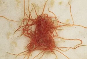
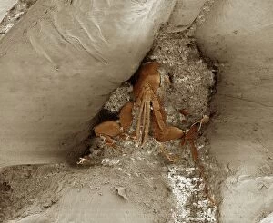
Picture No. 10855792Tubifex Worms - freshwater stream (Lumbriculus variegatus ) Date:
Marine DiatomsLRMC-3 Scanning Electron Micrograph (SEM): Marine Diatoms Magnification x2400 (when printed A4, 29.7 cm cm wide) David McCarthy and Dae Sasitorn / ardea.com Last Refuge contact details
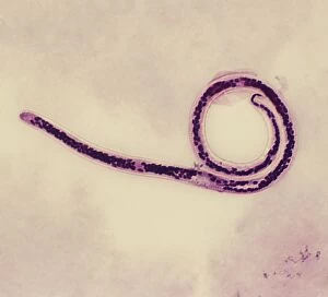
Light Micrograph (SEM): Micro-filarial worm - Magnification x 3000 (if print A4 size: 29. 7 cm wide)LRDS-314 Light Micrograph (SEM): Micro-filarial worm Magnification x 3000 (if print A4 size: 29.7 cm wide) Wuchereria bancrofti Elephantiasis David Spears (Last Refuge)
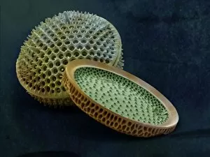
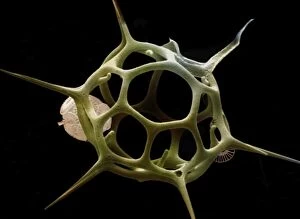
SilicoflagellateLRDS-297 Scanning Electron Micrograph (SEM): Silicoflagellate, Skeleton bearing stage of Dictyocha speculum Magnification x3
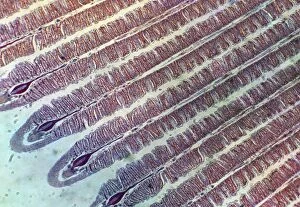
Microscopic Fish GillPM-1947 Fish Anatomy - Fish gill under microscope. Pat Morris Please note that prints are for personal display purposes only and may not be reproduced in anyway
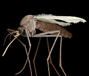
Scanning Electron Micrograph (SEM): Mosquito, Magnification x 35 (A4 size: 29. 7 cm width)LRDS-84 Mosquito Scanning Electron Micrograph (SEM) Culex pipiens Magnification x 35 (A4 size: 29.7 cm width) Coloured by hand to enhance natural features
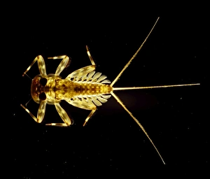
Light Micrograph : Flattened Mayfly Nymph, Magnification x 10 (A4 size: 29. 7 cm width)LRDS-80 Flattened Mayfly Nymph Light Micrograph Ecdyonurus venosus Magnification x 10 (A4 size: 29.7 cm width) Credit: David Spears (last refuge)
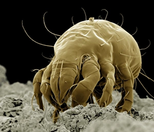
Scanning Electron Micrograph (SEM): Flour Mite, Magnification x 500 (A4 size: 29. 7 cm width)LRDS-58 Flour Mite Scanning Electron Micrograph (SEM) Acarus siro Magnification x 500 (A4 size: 29.7 cm width) Coloured by hand to enhance natural features
Scanning Electron Micrograph (SEM): Cat Flea, Magnification x250 (A4 size: 29. 7 cm width)LRDS-54 Cat Flea Scanning Electron Micrograph (SEM) Ctenocephalides felis Magnification x250 (A4 size: 29.7 cm width) Coloured by hand to enhance natural features
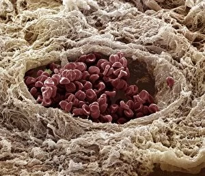
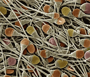
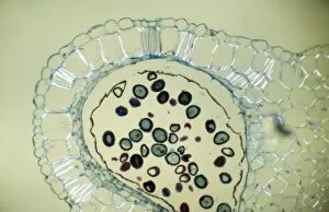
Light Micrograph (LM): Transverse section of Dehiscence Lilium Anthers with Pollen; Magnification x300 (on 10)LRDS-200 Light Micrograph (LM): Transverse section of Dehiscence Lilium Anthers with Pollen Magnification x300 (on 10.5 cm width print) David Spears (Last Refuge)
Scanning Electron Micrograph (SEM): Chigoe Flea, Magnification x 200 (A4 size: 29. 7 cm width)LRDS-137 Chigoe Flea Scanning Electron Micrograph (SEM) Tunga penetrans Magnification x 200 (A4 size: 29.7 cm width) Coloured by hand to enhance natural features
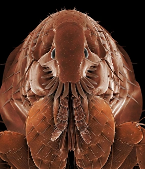
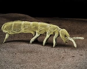
Scanning Electron Micrograph (SEM): Springtail, Order Collembola; Magnification x 150 (A4 size: 29. 7 cm width)LRDS-122 Springtail, Order Collembola Scanning Electron Micrograph (SEM) Magnification x 150 (A4 size: 29.7 cm width) Coloured by hand to enhance natural features