mail_outline sales@mediastorehouse.com
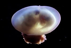
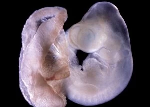
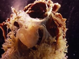
Picture No. 10875787Light Micrograph of Rat Embryo - 14.5 days into its gestation period - in sac with placenta Date:
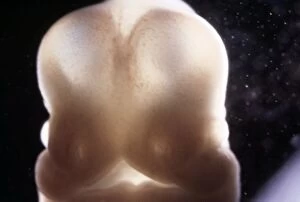
Picture No. 10875793Light Micrograph of Rat Embryo without its yolk sac, at 17.5 days Date:
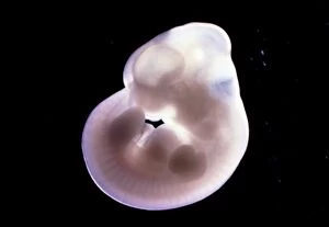
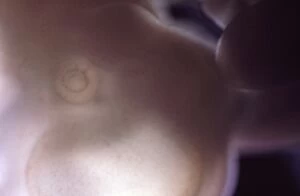
Picture No. 10875789Light Micrograph of Rat Embryo at 14.5days old Date:
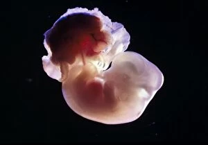
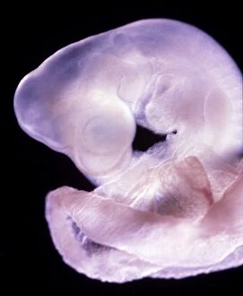
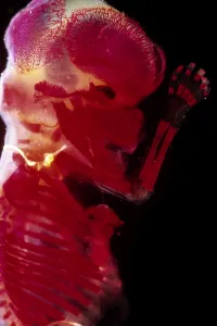
Picture No. 10875791Light Micrograph of Rat Embryo at 15.5 days, no sac, no placenta Date:
Picture No. 10875788Light Micrograph of Rat Embryo - 14.5 days into its gestation period - in sac with placenta Date:
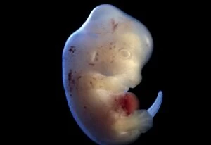
Picture No. 10875785Light Micrograph of Rat Embryo 13.2 days after fertilisation Date:
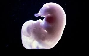
Picture No. 10875782Light Micrograph of Rat Embryo 11.5 days after fertilisation Date:
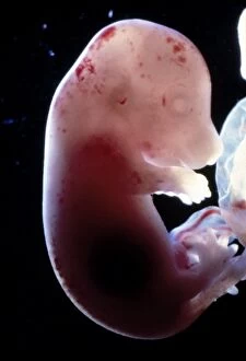
Picture No. 10875786Light Micrograph of Rat Embryo 13.2 days after fertilisation, out of its yolk sac Date:
Picture No. 10875780Light Micrograph of Rat Embryo 11.5 days after fertilisation Date:
Picture No. 10875783Light Micrograph of Rat Embryo 13.2 days after fertilisation Date:
Picture No. 10875784Light Micrograph of Eye detail of a rat embryo at 13.2 days after fertilisation Date:
Picture No. 10875781Light Micrograph of Rat Embryo 11.5 days after fertilisation Date:
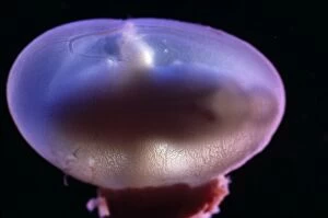
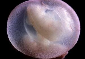
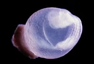
Picture No. 10875779Light Micrograph of Rat Embryo 10.2 days after fertilisation in sac Date:
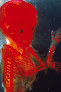
Picture No. 10875775Light Micrograph of Human Foetus 12 weeks - red dye to show bones Date:
Picture No. 10875778Light Micrograph of Human Embryo With Placenta Date:
Picture No. 10875776Light Micrograph of Human Foetus 12 weeks - red dye to show bones Date:
Picture No. 10875777Light Micrograph of Human Foetus 10-11 weeks after fertilisation Date:
Picture No. 10875774Light Micrograph of Human Foetus 12 weeks - red dye to show bones; Date:
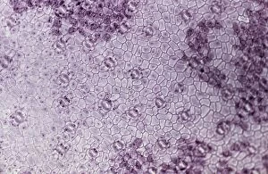
Picture No. 10874465Light Micrograph (LM): A transverse section of a lower Epidermis of a Box leaf (Buxus sp.) showing Stomata (Buxus sp.) Date:
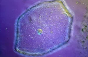
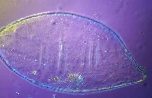
Picture No. 10874442Light Micrograph (LM): Tomato seed; Magnification x600 (on 10.5 cm width print) Date:
Picture No. 10874441Light Micrograph (LM): Tomato seed; Magnification x600 (on 10.5 cm width print) Date:
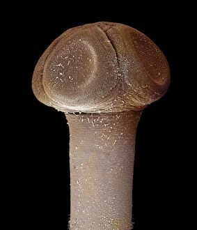
Picture No. 10874543Scanning Electron Micrograph (SEM): Tapeworm from a teleost fish Date:
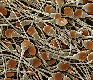
Picture No. 10874544Scanning Electron Micrograph (SEM): human sperm Date:
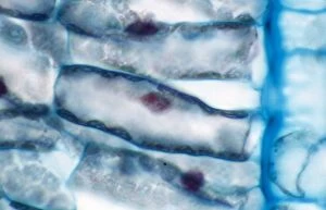
Picture No. 10874514Light Micrograph (LM): A light micrograph of the nuclei of plant cells Date:
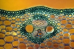
Picture No. 10874466Light Micrograph (LM): A transverse section of a Maize stem (Zea sp.) (Zea sp) Date:
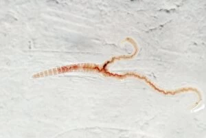
Picture No. 10855807Mutant Tiger Worm - juvenile with two tails (Eisenia fetida) Date:
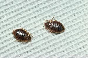
Picture No. 10851900BED BUGS - x 2 (Cimex lectularius) Date:
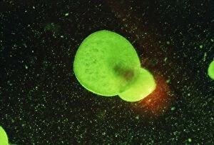
Picture No. 10851665Rootless / Least DUCKWEED (X5) (Wolffia arrhiza) Date:
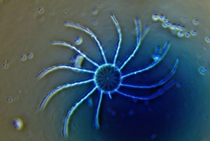
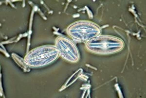
Diatom - from marine plankton sample - Hong Kong harbourSPH-552 Diatom - from marine plankton sample Hong Kong harbour Steve Hopkin Microscopic Please note that prints are for personal display purposes only and may not be reproduced in any way
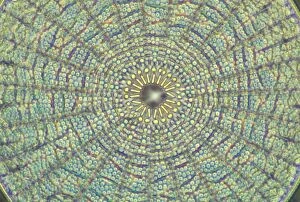
Diatoms - from marine plankton sample - Santa Anna - CaliforniaSPH-532 Diatoms - from marine plankton sample Santa Anna, California, USA Steve Hopkin Microscopic Please note that prints are for personal display purposes only and may not be reproduced in any way
Diatoms - from marine plankton sample - Hong Kong harbourSPH-534 Diatoms - from marine plankton sample Hong Kong harbour Steve Hopkin Microscopic Please note that prints are for personal display purposes only and may not be reproduced in any way
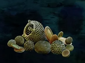
Marine Diatoms and RadiolariansLRMC-17 Scanning Electron Micrograph (SEM): Marine Diatoms and Radiolaria skeletons Magnification x435 (when printed A4, 29.7 cm cm wide)
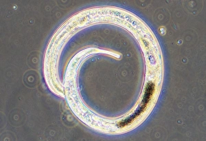
Microscopic File Nematode Worm From sediment of garden pond, also found inl film of water in soil, UKSPH-25 Microscopic - Nematode Worm from sediment of garden pond, also found in film of water in soil UK. Steve Hopkin contact details: prints@ardea.com tel: +44 (0) 20 8318 1401
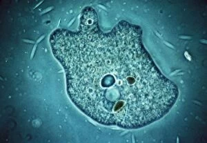
Amoeba - Phase contrastJC-183 AMOEBA - PHASE CONTRAST John Clegg contact details: prints@ardea.com tel: +44 (0) 20 8318 1401
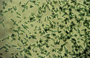
Protozoa - x 50 magnificationJC-202 Protozoa - x 50 magnification Euglena flagellata John Clegg contact details: prints@ardea.com tel: +44 (0) 20 8318 1401
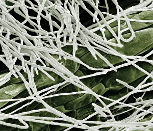
Scanning Electron Micrograph (SEM): Fusarium, Magnification x 1, 600 (A4 size: 29. 7 cm width)LRDS-60 Fusarium Scanning Electron Micrograph (SEM) Fusarium graminearum Magnification x 1, 600 (A4 size: 29.7 cm width) Coloured by hand to enhance natural features
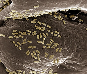
Scanning Electron Micrograph (SEM): E. coli; Magnification x 15, 000 (A4 size: 29. 7 cm width)LRDS-40 E. coli Scanning Electron Micrograph (SEM) Escherichia coli Magnification x 15, 000 (A4 size: 29.7 cm width) Coloured by hand to enhance natural features