mail_outline sales@mediastorehouse.com
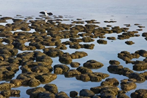
Stromatolites at Hamelin Pool, Shark Bay, western Australia. These are living representatives of the oldest livingROG-13733 Stromatolites at Hamelin Pool, Shark Bay, western Australia These are living representatives of the oldest living organisms, over 3.5 billion years old as fossils
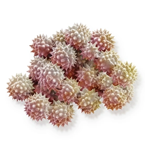
Scanning Electron Micrograph (SEM): Groundsel, Magnification x 3, 000 (A4 size: 29. 7 cm width)LRDS-98 Groundsel Scanning Electron Micrograph (SEM) Senecio vulgaris Magnification x 3, 000 (A4 size: 29.7 cm width) Coloured by hand to enhance natural features
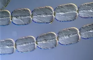
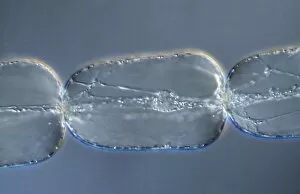
Light Micrograph (LM): Cells from a hair on the stamen of the common spiderwort (Tradescantia)LRDS-171 Light Micrograph (LM): Cells from a hair on the stamen of the common spiderwort (Tradescantia) Tradescantia sp David Spears (Last Refuge)
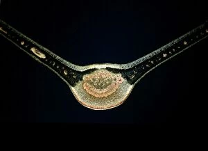
Light Micrograph (LM): A transverse section of a Fig leaf); Magnification x15 (on 10. 5 cm width print)LRDS-187 Light Micrograph (LM): A transverse section of a Fig leaf) Magnification x15 (on 10.5 cm width print) David Spears (Last Refuge)
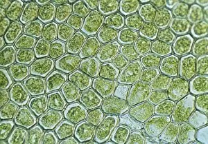
Light Micrograph (LM): The cellular struture of a liverwort plant (Hepatica); Magnification x1200 (on 10)LRDS-190 Light Micrograph (LM): The cellular struture of a liverwort plant (Hepatica) Magnification x1200 (on 10.5 cm width print) Hepatica sp David Spears (Last Refuge)
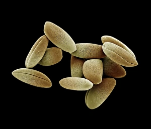
Scanning Electron Micrograph (SEM): Lily Pollen, Magnification x 1, 200 (A4 size: 29. 7 cm width) LRDS-99 Lily Pollen Scanning Electron Micrograph (SEM) Lilium sp. Magnification x 1, 200 (A4 size: 29.7 cm width) Coloured by hand to enhance natural features
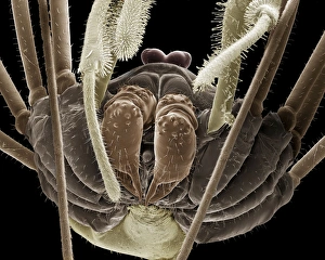
Scanning Electron Micrograph (SEM): Harvestman, Magnification x 70 (A4 size: 29. 7 cm width)LRDS-66 Harvestman Scanning Electron Micrograph (SEM) Phalangium opilio Magnification x 70 (A4 size: 29.7 cm width) Coloured by hand to enhance natural features
Light Micrograph (LM): A single cell from a hair on the stamen of the common spiderwort (Tradescantia)LRDS-170 Light Micrograph (LM): A single cell from a hair on the stamen of the common spiderwort (Tradescantia) Tradescantia sp David Spears (Last Refuge)
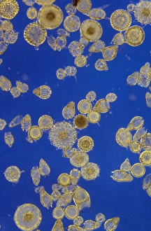
Protozoa E. MediterraneanJC-75 Protozoa E. Mediterranean Formaminifera ooze John Clegg Please note that prints are for personal display purposes only and may not be reproduced in any way
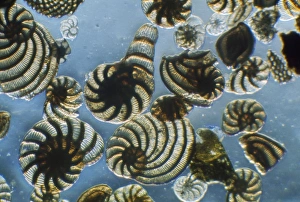
Protozoa - Radiolarians (x20) ActinopodaJC-291 Protozoa - Radiolarians (x20) Actinopoda John Clegg Please note that prints are for personal display purposes only and may not be reproduced in any way
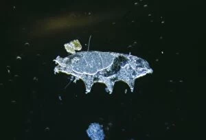
Microscopic Water-bear Freshwater arthropodJC-339 Freshwater TARDIGRADE - Water Bear / Tardigrada John Clegg Microscopic Please note that prints are for personal display purposes only and may not be reproduced in any way
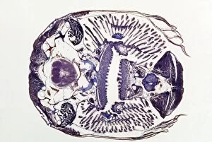
Microscopic Fish Gill - tranverse section through gill region of fish. Fish anatomyJC-429 Microscopic Fish Gill - tranverse section through gill region of fish. Fish anatomy. John Clegg Please note that prints are for personal display purposes only
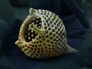
RadiolarianLRMC-13 Scanning Electron Micrograph (SEM): Radiolaria skeleton: Magnification x960 (when printed A4, 29.7 cm cm wide) Lychnocanomma bellum David McCarthy
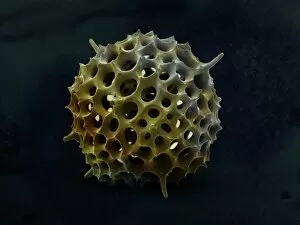
RadiolarianLRMC-12 Scanning Electron Micrograph (SEM): Spumellarian Radiolaria Magnification x1900 (when printed A4, 29.7 cm cm wide)
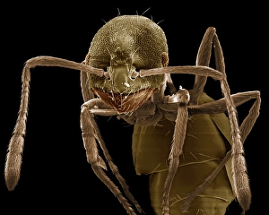
Scanning Electron Micrograph (SEM): Pharaoh Ant, Magnification x 180 (A4 size: 29. 7 cm width)LRDS-91 Pharaoh Ant Scanning Electron Micrograph (SEM) Monomorium pharaonis Magnification x 180 (A4 size: 29.7 cm width) Coloured by hand to enhance natural features
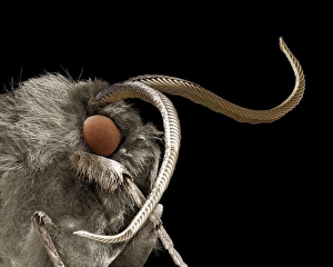
Scanning Electron Micrograph (SEM): Footman moth, Family Arctiidae ; Magnification x 25 (A4 size: 29. 7 cm width)LRDS-79 Footman moth, Family Arctiidae Scanning Electron Micrograph (SEM) Magnification x 25 (A4 size: 29.7 cm width) Coloured by hand to enhance natural features
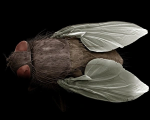
Scanning Electron Micrograph (SEM): House Fly, Magnification x 25 (A4 size: 29. 7 cm width)LRDS-71 House Fly, Scanning Electron Micrograph (SEM) Musca domestica Magnification x 25 (A4 size: 29.7 cm width) Coloured by hand to enhance natural features
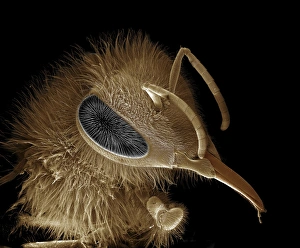
Scanning Electron Micrograph (SEM): Honeybee, Magnification x 30 (A4 size: 29. 7 cm width)LRDS-70 Honeybee Scanning Electron Micrograph (SEM) Apis mellifera Magnification x 30 (A4 size: 29.7 cm width) Coloured by hand to enhance natural features
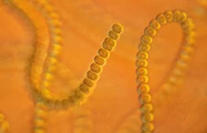
Light Micrograph: Cyanobacterium. ; Magnification x 3, 000 (A4 size: 29. 7 cm width)LRDS-7 Cyanobacterium Light Micrograph Anabaena sp Magnification x 3, 000 (A4 size: 29.7 cm width) Credit: David Spears (last refuge)
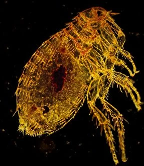
Oriental Rat Flea, Magnification x 200 (A4 size: 29. 7 cm width)LRDS-53 Oriental Rat Flea Dark Field Light Micrograph (LM) Xenopsylla cheopis Magnification x 200 (A4 size: 29.7 cm width) Credit
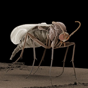
Scanning Electron Micrograph (SEM): Fungus Gnat, ; Magnification x 70 (A4 size: 29. 7 cm width)LRDS-50 Fungus Gnat Scanning Electron Micrograph (SEM) Bradysia paupera Magnification x 70 (A4 size: 29.7 cm width) Coloured by hand to enhance natural features
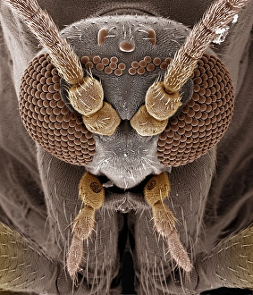
Scanning Electron Micrograph (SEM): Fungus Gnat, ; Magnification x 350 (A4 size: 29. 7 cm width)LRDS-49 Fungus Gnat Scanning Electron Micrograph (SEM) Bradysia paupera Magnification x 350 (A4 size: 29.7 cm width) Coloured by hand to enhance natural features
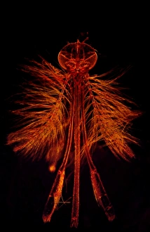
Dark Field Light Micrograph: Mosquito - male. ; Magnification x 65 (A4 size: 29. 7 cm width)LRDS-4 Mosquito - male Dark Field Light Micrograph (LM) Anopheles sp Magnification x 65 (A4 size: 29.7 cm width) Credit: David Spears (last refuge)