mail_outline sales@mediastorehouse.com
301 items
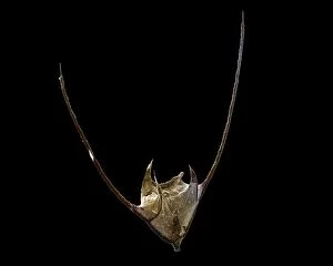
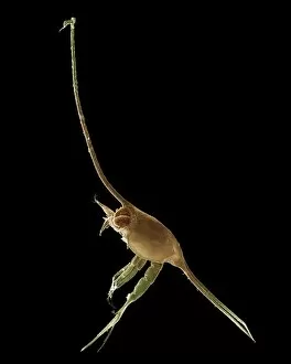
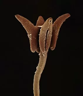
Scanning Electron Micrograph (SEM): Dinoflagellate, Ceratium sp. ; Magnification x 410 (when printed A4, 29. 7 cm wide)LRDS-275 Scanning Electron Micrograph (SEM): Dinoflagellate, Ceratium sp Magnification x 410 (when printed A4, 29.7 cm wide) Ceratium sp David Spears (Last Refuge) / ardea.com Last Refuge
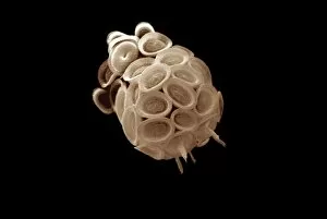
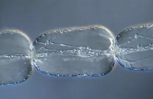
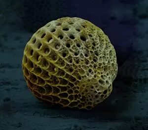
CoccolithophoreLRDS-280 Scanning Electron Micrograph (SEM): Coccolithophore (Calcareous Nannoplankton) Magnification x2, 250 (when printed 10.5 cm wide) Syracosphaera pulchra David Spears (Last Refuge)
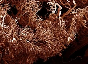
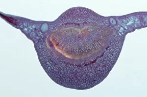
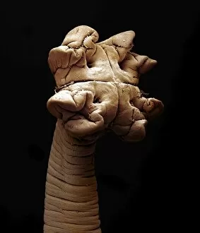
Scanning Electron Micrograph (SEM): a corrosion cast of a gut tumour. Liquid plastic is injected into the bloodLRDS-273 Scanning Electron Micrograph (SEM): a corrosion cast of a gut tumour Liquid plastic is injected into the blood vessels; it solidifies
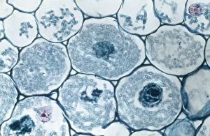
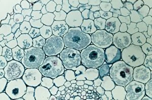
Light Micrograph (LM): Filaments of fungi Endotrophic Mycorrhiza live within cells of a root; Magnification x1200LRDS-253 Light Micrograph (LM): Filaments of fungi Endotrophic Mycorrhiza live within cells of a root Magnification x1200 (on 10.5 cm width print) David Spears (Last Refuge) / ardea.com Last Refuge
Light Micrograph (LM): Filaments of fungi Endotrophic Mycorrhiza live within cells of a root; Magnification x600LRDS-254 Light Micrograph (LM): Filaments of fungi Endotrophic Mycorrhiza live within cells of a root Magnification x600 (on 10.5 cm width print) David Spears (Last Refuge) / ardea.com Last Refuge
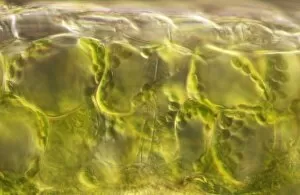
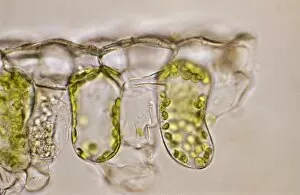
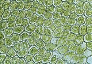
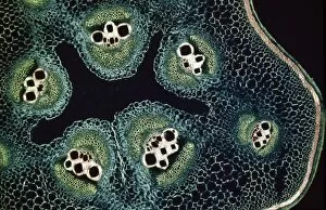
Light Micrograph (LM): plant cell chloroplasts - the site where photosynthesis takes placeLRDS-192 Light Micrograph (LM): plant cell chloroplasts - the site where photosynthesis takes place David Spears (Last Refuge)
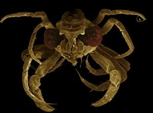
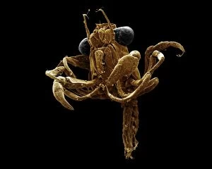
Scanning Electron Micrograph (SEM): Megalop; Magnification x 90 (when printed A4, 29. 7 cm wide)LRDS-270 Scanning Electron Micrograph (SEM): Megalop Magnification x 90 (when printed A4, 29.7 cm wide) David Spears (Last Refuge)
Light Micrograph (LM): plant cell chloroplasts - the site where photosynthesis takes placeLRDS-191 Light Micrograph (LM): plant cell chloroplasts - the site where photosynthesis takes place David Spears (Last Refuge)
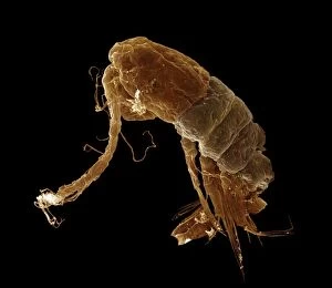
Scanning Electron Micrograph (SEM): Calanus sp. ; Magnification x650 (A4 size: 29. 7 cm width)LRDS-302 Scanning Electron Micrograph (SEM): Calanus sp Magnification x650 (A4 size: 29.7 cm width) David Spears (Last Refuge)
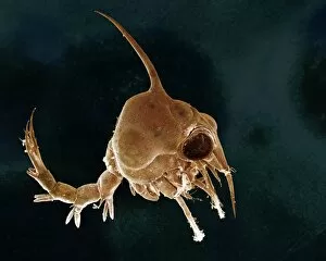
Scanning Electron Micrograph (SEM): Crab larvae (Zoea larvae); Magnification x 70 (when printed A4, 29. 7 cm wide)LRDS-263 Scanning Electron Micrograph (SEM): Crab larvae (Zoea larvae) Magnification x 70 (when printed A4, 29.7 cm wide) The zoea is a free-swimming larval stage of crustaceans
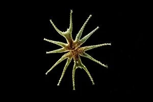
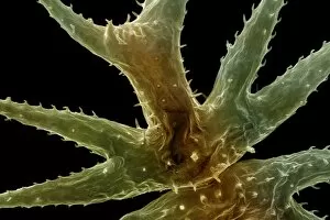
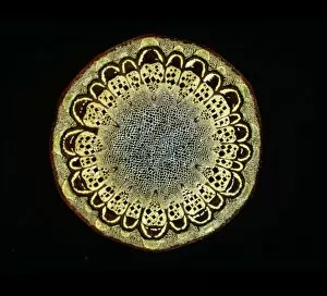
Freshwater DesmidLRDS-303 Scanning Electron Micrograph (SEM): Freshwater Desmid Magnification x260 (when printed 10.5 cm Micrasterias lux David Spears (Last Refuge)
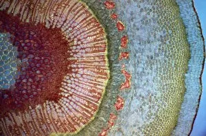
Light Micrograph (LM): A transverse section of a leaf of Tasmanian Blue Gum (Eucalyptus globulus)LRDS-186 Light Micrograph (LM): A transverse section of a leaf of Tasmanian Blue Gum (Eucalyptus globulus) Magnification x30 (on 10.5 cm width print) Eucalyptus globulus David Spears (Last Refuge)
Light Micrograph (LM): Cells from a hair on the stamen of the common spiderwort (Tradescantia)LRDS-171 Light Micrograph (LM): Cells from a hair on the stamen of the common spiderwort (Tradescantia) Tradescantia sp David Spears (Last Refuge)
Light Micrograph (LM): A transverse section of a Fig leaf); Magnification x15 (on 10. 5 cm width print)LRDS-187 Light Micrograph (LM): A transverse section of a Fig leaf) Magnification x15 (on 10.5 cm width print) David Spears (Last Refuge)
Freshwater DesmidLRDS-304 Scanning Electron Micrograph (SEM): Freshwater Desmid Magnification x1050 (when printed 10.5 cm Micrasterias lux David Spears (Last Refuge)
Light Micrograph (LM): The cellular struture of a liverwort plant (Hepatica); Magnification x1200 (on 10)LRDS-190 Light Micrograph (LM): The cellular struture of a liverwort plant (Hepatica) Magnification x1200 (on 10.5 cm width print) Hepatica sp David Spears (Last Refuge)
Scanning Electron Micrograph (SEM): Crab larvae (Zoea larvae); Magnification x 70 (when printed A4, 29. 7 cm wide)LRDS-272 Scanning Electron Micrograph (SEM): Crab larvae (Zoea larvae) Magnification x 70 (when printed A4, 29.7 cm wide) The zoea is a free-swimming larval stage of crustaceans
Scanning Electron Micrograph (SEM): Megalop; Magnification x 70 (when printed A4, 29. 7 cm wide)LRDS-271 Scanning Electron Micrograph (SEM): Megalop Magnification x 70 (when printed A4, 29.7 cm wide) David Spears (Last Refuge)
Light Micrograph (LM): A single cell from a hair on the stamen of the common spiderwort (Tradescantia)LRDS-170 Light Micrograph (LM): A single cell from a hair on the stamen of the common spiderwort (Tradescantia) Tradescantia sp David Spears (Last Refuge)
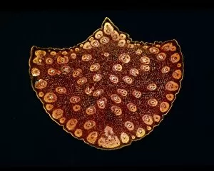
Light Micrograph (LM): A transverse section of a stem of a Marrow (Cucurbita sp. ); Magnification x12 (on 10)LRDS-193 Light Micrograph (LM): A transverse section of a stem of a Marrow (Cucurbita sp.) Magnification x12 (on 10.5 cm width print) Cucurbita sp
Light Micrograph (LM): A transverse section of a stem of Jerusalem artichoke (Helianthus tuberosus)LRDS-166 Light Micrograph (LM): A transverse section of a stem of Jerusalem artichoke (Helianthus tuberosus) Magnification x600 (on 10.5 cm width print) Helianthus tuberosus David Spears (Last Refuge)
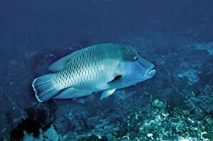
Maori / Napoleon Wrasse - female - IndonesiaVT-8928 Maori / Napoleon Wrasse - female Indonesia Cheilinus undulatus Endangered Valerie & Ron Taylor Please note that prints are for personal display purposes only
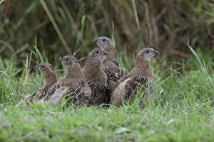
Grey Partridge - five Juveniles huddled together beside hedge bottom - July - Gooderstone Norfolk UKCK-4459 Grey Partridge - five Juveniles huddled together beside hedge bottom July - Gooderstone Norfolk UK Perdix perdix Chris Knights Please note that prints are for personal display purposes only
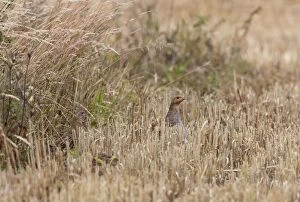
Grey Partridge - female on edge of barley stubble field with young - July - Gooderstone Noroflk UKCK-4458 Grey Partridge - female on edge of barley stubble field with young July - Gooderstone Norfolk UK Perdix perdix Chris Knights Please note that prints are for personal display purposes only
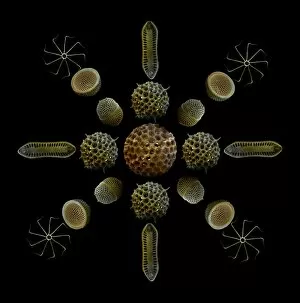
Scanning Electron Micrograph (SEM): Marine Diatoms and Radiolaria skeletons; Magnification x435 (when printed A4, 29)LRMC-14 Scanning Electron Micrograph (SEM): Marine Diatoms and Radiolaria skeletons Magnification x435 (when printed A4, 29.7 cm cm wide)
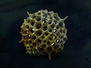
RadiolarianLRMC-13 Scanning Electron Micrograph (SEM): Radiolaria skeleton: Magnification x960 (when printed A4, 29.7 cm cm wide) Lychnocanomma bellum David McCarthy
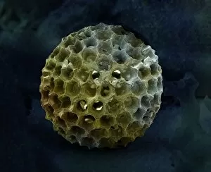
RadiolarianLRMC-12 Scanning Electron Micrograph (SEM): Spumellarian Radiolaria Magnification x1900 (when printed A4, 29.7 cm cm wide)
RadiolarianLRMC-11 Scanning Electron Micrograph (SEM): Spumellarian Radiolaria Magnification x1450 (when printed A4, 29.7 cm cm wide)
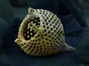
RadiolarianLRMC-10 Scanning Electron Micrograph (SEM): Radiolaria skeleton, Zamoidellum ovum or Carpocanistrum favosum Magnification x2400 (when printed A4)
Scanning Electron Micrograph (SEM): Tapeworm from sharkLRDS-265 Scanning Electron Micrograph (SEM): Tapeworm from shark David Spears (Last Refuge) / ardea.com Last Refuge Please note that prints are for personal display purposes only
Scanning Electron Micrograph (SEM): Tapeworm from sharkLRDS-264 Scanning Electron Micrograph (SEM): Tapeworm from shark David Spears (Last Refuge) / ardea.com Last Refuge Please note that prints are for personal display purposes only
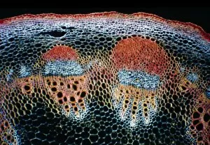
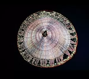
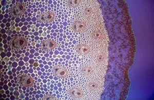
Light Micrograph (LM): A transverse section of a stem of Fragrant Virgin's Bower (Clematis flammula)LRDS-262 Light Micrograph (LM): A transverse section of a stem of Fragrant Virgin's Bower stem Magnification x30 (on 10.5 cm width print) Clematis flammula David Spears (Last Refuge)
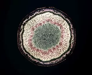
Light Micrograph (LM): A transverse section of a stem of Common Lime tree (Tilia europaea); Magnification x12 (on 10)LRDS-261 Light Micrograph (LM): A transverse section of a stem of Common Lime tree Magnification x12 (on 10.5 cm width print) Tilia europaea David Spears (Last Refuge)
Light Micrograph (LM): Transverse section of a stem of Sycamore (Acer pseudoplatanus); Magnification x12 (on 10)LRDS-260 Light Micrograph (LM): Transverse section of a stem of Sycamore Magnification x12 (on 10.5 cm width print) Acer pseudoplatanus David Spears (Last Refuge)
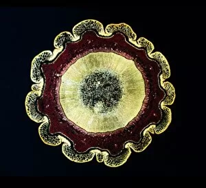
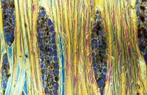
Light Micrograph (LM): The transverse section of a stem of Norway Pine (Abies Excelsa)LRDS-259 Light Micrograph (LM): The transverse section of a stem of Norway Pine shows the resin passages Magnification x24 (on 10.5 cm width print) Abies Excelsa David Spears (Last Refuge)
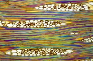
Light Micrograph (LM): A Tranverse section of a stem of a Common or European Ash tree (Fraxinus excelsior)LRDS-258 Light Micrograph (LM): A Tranverse section of a stem of a Common or European Ash tree Magnification x12 (on 10.5 cm width print) Fraxinus excelsior David Spears (Last Refuge)
Light Micrograph (LM): A Tranverse section of a stem of a Common or European Ash tree (Fraxinus excelsior)LRDS-257 Light Micrograph (LM): A Tranverse section of a stem of a Common or European Ash tree Magnification x30 (on 10.5 cm width print) Fraxinus excelsior David Spears (Last Refuge)
Light Micrograph (LM): A transverse section of a stem of a Palm; Magnification x12 (on 10. 5 cm width print)LRDS-256 Light Micrograph (LM): A transverse section of a stem of a Palm Magnification x12 (on 10.5 cm width print) David Spears (Last Refuge)
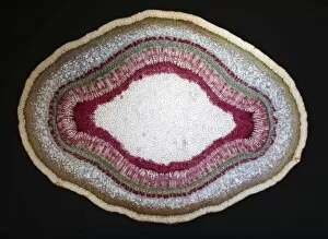
Light Micrograph (LM): A transverse section of a stem of a Butcher's broom (Ruscus aculeatus)LRDS-255 Light Micrograph (LM): A transverse section of a stem of a Butcher's broom Magnification x150 (on 10.5 cm width print) Ruscus aculeatus David Spears (Last Refuge)
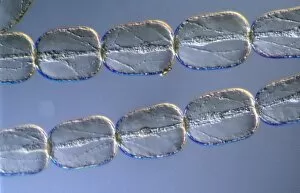
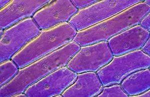
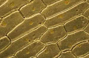
Light Micrograph (LM): Onion skin cells; Magnification x600 (on 10. 5 cm width print)LRDS-252 Light Micrograph (LM): Onion skin cells; Magnification x600 (on 10.5 cm width print) David Spears (Last Refuge) / ardea.com Last Refuge Please note that prints are for personal display
Light Micrograph (LM): Onion skin cells; Magnification x600 (on 10. 5 cm width print)LRDS-251 Light Micrograph (LM): Onion skin cells; Magnification x600 (on 10.5 cm width print) David Spears (Last Refuge) / ardea.com Last Refuge Please note that prints are for personal display
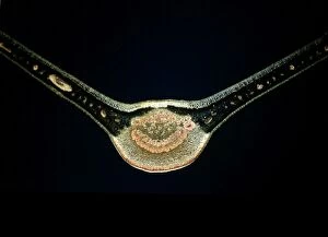
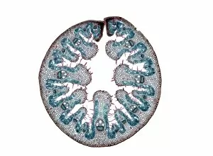
Light Micrograph (LM): Transverse section of a leaf of Marram Grass (Ammophila sp. ); Magnification x15 (on 10)LRDS-250 Light Micrograph (LM): Transverse section of a leaf of Marram Grass Magnification x15 (on 10.5 cm width print) Ammophila sp
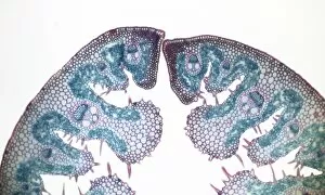
Light Micrograph (LM): Transverse section of a leaf of Marram Grass (Ammophila sp. ); Magnification x300 (on 10)LRDS-249 Light Micrograph (LM): Transverse section of a leaf of Marram Grass Magnification x300 (on 10.5 cm width print) Ammophila sp
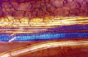
Light Micrograph (LM): Longitudinal section of stem of Crown of Thorns (Euphorbia splendens)LRDS-248 Light Micrograph (LM): Longitudinal section of stem of Crown of Thorns Magnification x1200 (on 10.5 cm width print) Euphorbia splendens David Spears (Last Refuge)
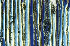
Light Micrograph (LM): Longitudinal section of Mahogany wood, (Pinus sylvestris); Magnification x600 (on 10)LRDS-247 Light Micrograph (LM): Longitudinal section of Mahogany wood, Magnification x600 (on 10.5 cm width print) Pinus sylvestris David Spears (Last Refuge)
Light Micrograph (LM): Longitudinal section of Mahogani (Swietenia mahagoni); Magnification x600 (on 10)LRDS-246 Light Micrograph (LM): Longitudinal section of Mahogani Magnification x600 (on 10.5 cm width print) Swietenia mahagoni David Spears (Last Refuge)
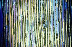
Light Micrograph (LM): Longitudinal section of Scots pine wood, (Pinus sylvestris); Magnification x480 (on 10)LRDS-245 Light Micrograph (LM): Longitudinal section of Scots pine wood, Magnification x480 (on 10.5 cm width print) Pinus sylvestris David Spears (Last Refuge)
Light Micrograph (LM): Longitudinal section of Scots pine wood, (Pinus sylvestris); Magnification x600 (on 10)LRDS-244 Light Micrograph (LM): Longitudinal section of Scots pine wood Magnification x600 (on 10.5 cm width print) Pinus sylvestris David Spears (Last Refuge)