mail_outline sales@mediastorehouse.com
301 items
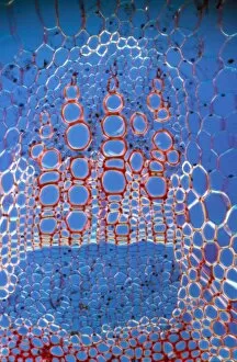
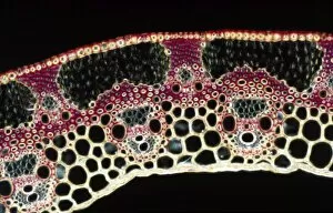
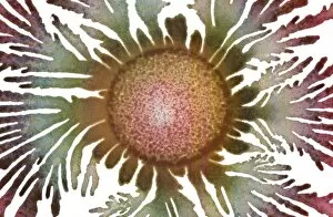
Light Micrograph (LM): Transverse section shows Sclerenchyma Ground Tissue in Helianthus stem; Magnification x600LRDS-243 Light Micrograph (LM): Transverse section shows Sclerenchyma Ground Tissue in Helianthus stem Magnification x600 (on 10.5 cm width print) Helianthus sp David Spears (Last Refuge)
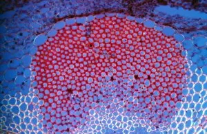
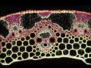
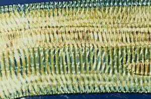
Light Micrograph (LM): A transverse section shows Xylem in Helianthus stem; Magnification x600 (on 10)LRDS-242 Light Micrograph (LM): A transverse section shows Xylem in Helianthus stem Magnification x600 (on 10.5 cm width print) Helianthus sp David Spears (Last Refuge)
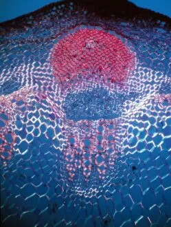
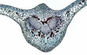
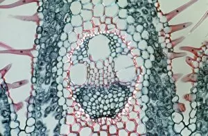
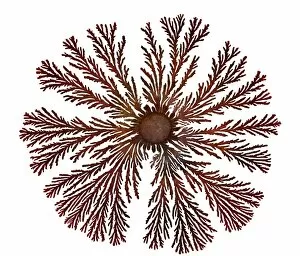
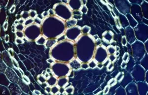
Light Micrograph (LM): A transverse section shows Vascular Bundle in Helianthus stem; Magnification x300 (on 10)LRDS-241 Light Micrograph (LM): A transverse section shows Vascular Bundle in Helianthus stem Magnification x300 (on 10.5 cm width print) Helianthus sp David Spears (Last Refuge)
Light Micrograph (LM): A transverse section of a root of Creeping Buttercup, (Ranunculus repens)LRDS-240 Light Micrograph (LM): A transverse section of a root of Creeping Buttercup, Ranunculus repens David Spears (Last Refuge)
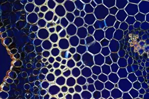
Light Micrograph (LM): A transverse section of a straw of Wheat; Magnification x600 (on 10. 5 cm width print)LRDS-239 Light Micrograph (LM): A transverse section of a straw of Wheat Magnification x600 (on 10.5 cm width print) David Spears (Last Refuge)
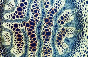
Light Micrograph (LM): A transverse section of a straw of Wheat; Magnification x1200 (on 10. 5 cm width print)LRDS-238 Light Micrograph (LM): A transverse section of a straw of Wheat Magnification x1200 (on 10.5 cm width print) David Spears (Last Refuge)
Light Micrograph (LM): A transverse section of a leaf of a Cherry (Prunus sp. ); Magnification x30 (on 10)LRDS-236 Light Micrograph (LM): A transverse section of a leaf of a Cherry Magnification x30 (on 10.5 cm width Prunus sp. David Spears (Last Refuge)
Light Micrograph (LM): Transverse section of a leaf of Marram Grass (Ammophila sp)LRDS-235 Light Micrograph (LM): Transverse section of a leaf of Marram Grass showing Vascular Bundle Magnification x1200 (on 10.5 cm width print) Ammophila sp
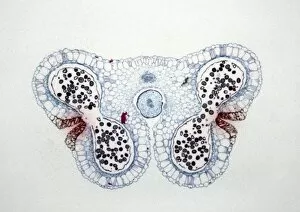
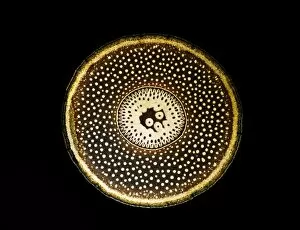
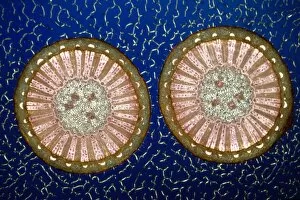
Light Micrograph (LM): Transverse section of Lilium Anthers with Mature Pollen; Magnification x15 (on 10)LRDS-234 Light Micrograph (LM): Transverse section of Lilium Anthers with Mature Pollen Magnification x15 (on 10.5 cm width print) David Spears (Last Refuge)
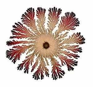
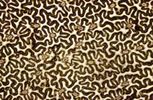
Macro Photograph: Patterns of Paenibacillus bacteria on petri dish - T type (tip-splitting morphotype)LRDS-233 Macro Photograph: Patterns of Paenibacillus bacteria on petri dish - T type (tip-splitting morphotype) magnification x5.6 (A4 size)
Macro Photograph: Patterns of Paenibacillus bacteria on petri dish - T type (tip-splitting morphotype)LRDS-230 Macro Photograph: Patterns of Paenibacillus bacteria on petri dish - T type (tip-splitting morphotype) magnification x5.6 (A4 size)
Macro Photograph: Patterns of Paenibacillus bacteria on petri dish - T type (tip-splitting morphotype)LRDS-229 Macro Photograph: Patterns of Paenibacillus bacteria on petri dish - T type (tip-splitting morphotype) magnification x5.6 (A4 size)
Macro Photograph: Patterns of Paenibacillus bacteria on petri dish - T type (tip-splitting morphotype)LRDS-228 Macro Photograph: Patterns of Paenibacillus bacteria on petri dish - T type (tip-splitting morphotype) magnification x5.6 (A4 size)
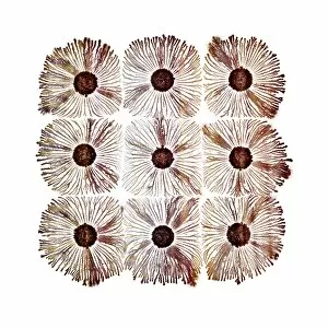
Macro Photograph: Patterns of Paenibacillus bacteria on petri dish - T type (tip-splitting morphotype)LRDS-227 Macro Photograph: Patterns of Paenibacillus bacteria on petri dish - T type (tip-splitting morphotype) magnification x5 (A4 size)
Macro Photograph: Patterns of Paenibacillus bacteria on petri dish - T type (tip-splitting morphotype)LRDS-226 Macro Photograph: Patterns of Paenibacillus bacteria on petri dish - T type (tip-splitting morphotype) magnification x5 (A4 size)
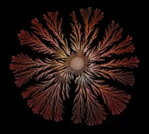
Macro Photograph: Patterns of Paenibacillus bacteria on petri dish - The colony structures form as adaptive responsesLRDS-225 Macro Photograph: Patterns of Paenibacillus bacteria on petri dish - The colony structures form as adaptive responses to laboratory-imposed stresses that mimic hostile environments faced in
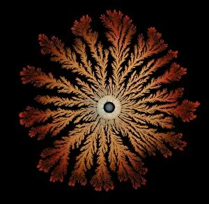
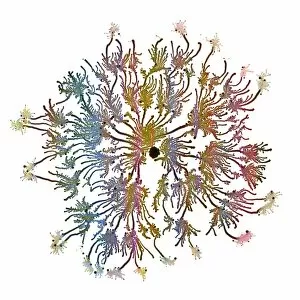
Macro Photograph: Patterns of Paenibacillus bacteria on petri dish - V type (vortex morphotype)LRDS-224 Macro Photograph: Patterns of Paenibacillus bacteria on petri dish - V type (vortex morphotype) magnification x5.5 (A4 size)
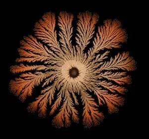
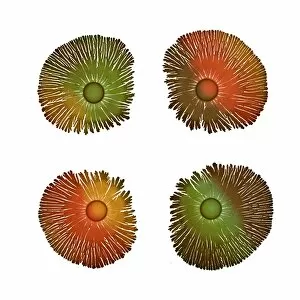
Macro Photograph: Patterns of Paenibacillus bacteria on petri dish - C type (chiral)LRDS-223 Macro Photograph: Patterns of Paenibacillus bacteria on petri dish - C type (chiral) magnification x5.5 (A4 size)
Macro Photograph: Patterns of Paenibacillus bacteria on petri dish - V type (vortex morphotype)LRDS-222 Macro Photograph: Patterns of Paenibacillus bacteria on petri dish - V type (vortex morphotype) magnification x5.5 (A4 size)
Macro Photograph: Patterns of Paenibacillus bacteria on petri dish - V type (vortex morphotype)LRDS-221 Macro Photograph: Patterns of Paenibacillus bacteria on petri dish - V type (vortex morphotype) magnification x5.6 (A4 size)
Macro Photograph: Patterns of Paenibacillus bacteria on petri dish - V type (vortex morphotype)LRDS-220 Macro Photograph: Patterns of Paenibacillus bacteria on petri dish - V type (vortex morphotype) magnification x5.5 (A4 size)
Macro Photograph: Patterns of Paenibacillus bacteria on petri dish - T type (tip-splitting morphotype)LRDS-219 Macro Photograph: Patterns of Paenibacillus bacteria on petri dish - T type (tip-splitting morphotype) magnification x30 (A4 size)
Macro Photograph: Patterns of Paenibacillus bacteria on petri dish - T type (tip-splitting morphotype)LRDS-217 Macro Photograph: Patterns of Paenibacillus bacteria on petri dish - T type (tip-splitting morphotype) magnification x25 (A4 size)
Macro Photograph: Patterns of Paenibacillus bacteria on petri dish - T type (tip-splitting morphotype)LRDS-216 Macro Photograph: Patterns of Paenibacillus bacteria on petri dish - T type (tip-splitting morphotype) magnification x5.6 (A4 size)
Macro Photograph: Patterns of Paenibacillus bacteria on petri dish - The colony structures form as adaptive responsesLRDS-215 Macro Photograph: Patterns of Paenibacillus bacteria on petri dish magnification x5 (A4 size: 29.7 cm width) The colony structures form as adaptive responses to laboratory-imposed stresses
Macro Photograph: Patterns of Paenibacillus bacteria on petri dish - C type (chiral)LRDS-214 Macro Photograph: Patterns of Paenibacillus bacteria on petri dish - C type (chiral) magnification x5.5 (A4 size)
Light Micrograph (LM): A transverse section of a stem of a Tree Fern (Dicksonia antractica); Magnification x9 (on 10)LRDS-213 Light Micrograph (LM): A transverse section of a stem of a Tree Fern Magnification x9 (on 10.5 cm width print) Dicksonia antractica David Spears (Last Refuge)
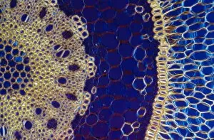
Light Micrograph (LM): A transverse section of a stem of a Monocot (Ruscus aculeatus); Magnification x30 (on 10)LRDS-212 Light Micrograph (LM): A transverse section of a stem of a Monocot Magnification x30 (on 10.5 cm width print) Ruscus aculeatus David Spears (Last Refuge)
Light Micrograph (LM): A transverse section of an aerial root from a Pandanus sp; Magnification x30 (on 10)LRDS-211 Light Micrograph (LM): A transverse section of an aerial root from a Pandanus sp Magnification x30 (on 10.5 cm width print) Pandanus sp David Spears (Last Refuge)
Light Micrograph (LM): A transverse section of a root of Conifer (Pandanus sp. ); Magnification x30 (on 10)LRDS-210 Light Micrograph (LM): A transverse section of a root of Conifer Magnification x30 (on 10.5 cm width print) Pandanus sp
Light Micrograph (LM): A transverse section of an aerial root of Orchid (Dendrobium sp. ); Magnification x30 (on 10)LRDS-209 Light Micrograph (LM): A transverse section of an aerial root of Orchid Magnification x30 (on 10.5 cm width print) Dendrobium sp
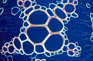
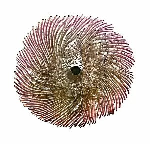
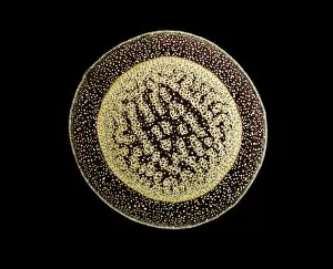
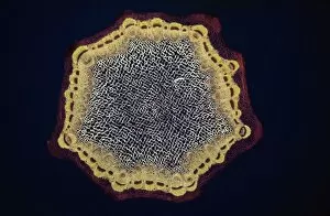
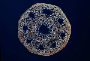
Light Micrograph (LM): A transverse section of a Helianthus stem; Magnification x18 (on 10. 5 cm width print)LRDS-208 Light Micrograph (LM): A transverse section of a Helianthus stem Magnification x18 (on 10.5 cm width print) Helianthus sp David Spears (Last Refuge)
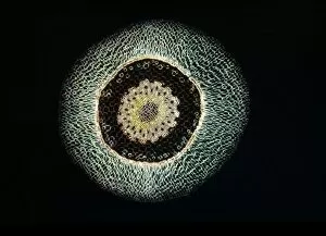
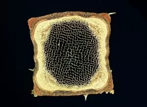
Light Micrograph (LM): A transverse section of a Dandelion stem (Taraxacum officinale); Magnification x24 (on 10)LRDS-207 Light Micrograph (LM): A transverse section of a Dandelion stem Magnification x24 (on 10.5 cm width print) Taraxacum officinale David Spears (Last Refuge)
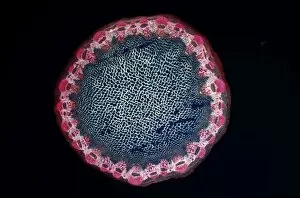
Light Micrograph (LM): A transverse section of a stem of a Pepper; Magnification x12 (on 10. 5 cm width print)LRDS-206 Light Micrograph (LM): A transverse section of a stem of a Pepper Magnification x12 (on 10.5 cm width print) David Spears (Last Refuge)
Light Micrograph (LM): A transverse section of a stem of a Hedge Woundwort plant (Stachys sylvatica)LRDS-205 Light Micrograph (LM): A transverse section of a stem of a Hedge Woundwort plant Magnification x60 (on 10.5 cm width print) Stachys sylvatica David Spears (Last Refuge)
Light Micrograph (LM): A transverse section of an aerial root of Orchid (Dendrobium sp. ); Magnification x600 (on 10)LRDS-204 Light Micrograph (LM): A transverse section of an aerial root of Orchid (Dendrobium sp.) Magnification x600 (on 10.5 cm width print) Dendrobium sp
Light Micrograph (LM): A transverse section of a stem of a Horsetail Fern (Equisetum sp. )LRDS-203 Light Micrograph (LM): A transverse section of a stem of a Horsetail Fern (Equisetum sp.) Equisetum sp. David Spears (Last Refuge)
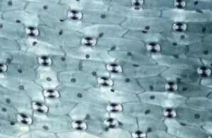
Light Micrograph (LM): A transverse section of a leaf of a Tulip (Tulipa sp)LRDS-202 Light Micrograph (LM): A transverse section of a leaf of a Tulip (Tulipa sp.) showing Stomata Magnification x600 (on 10.5 cm width print) Tulipa sp
Light Micrograph (LM): A transverse section of a leaf of a Tulip (Tulipa sp)LRDS-201 Light Micrograph (LM): A transverse section of a leaf of a Tulip (Tulipa sp.) showing Stomata Magnification x1200 (on 10.5 cm width print) Tulipa sp
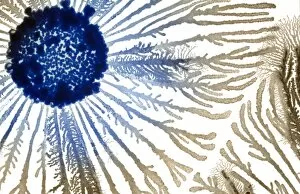
Light Micrograph (LM): Longitudinal section of old sunflower stem(Helianthus sp)LRDS-199 Light Micrograph (LM): Longitudinal section of old sunflower stem(Helianthus sp.) showing spiral chloroplasts Magnification x1200 (on 10.5 cm width print)
Light Micrograph (LM): Longitudinal section of old sunflower stem(Helianthus sp)LRDS-198 Light Micrograph (LM): Longitudinal section of old sunflower stem (Helianthus sp.) showing spiral chloroplasts Magnification x600 (on 10.5 cm width print)
Light Micrograph (LM): A concentration of cells on the epidermis of a plant showing stomataLRDS-197 Light Micrograph (LM): A concentration of cells on the epidermis of a plant showing stomata David Spears (Last Refuge)
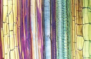
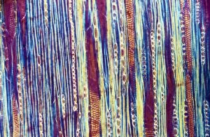
Light Micrograph (LM): Longitudinal section shows Scalariform vessels of Fern; Magnification x1200 (on 10)LRDS-196 Light Micrograph (LM): Longitudinal section shows Scalariform vessels of Fern Magnification x1200 (on 10.5 cm width print) David Spears (Last Refuge)
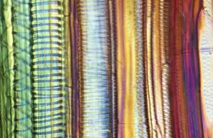
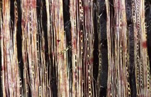
Light Micrograph (LM): A longitudinal section f a Ribes sp. stem; Magnification x600 (on 10. 5 cm width print)LRDS-195 Light Micrograph (LM): A longitudinal section of a Ribes sp. stem Magnification x600 (on 10.5 cm width print) Ribes sp
Light Micrograph (LM): A longitudinal section f a Ribes sp. stem; Magnification x600 (on 10. 5 cm width print)LRDS-194 Light Micrograph (LM): A longitudinal section of a Ribes sp. stem Magnification x600 (on 10.5 cm width print) Ribes sp
Light Micrograph (LM): A transverse section of a stem of Clubmoss(Lycopodium sp. ); Magnification x600 (on 10)LRDS-185 Light Micrograph (LM): A transverse section of a stem of Clubmoss (Lycopodium sp.) Magnification x600 (on 10.5 cm width print) Lycopodium sp
Light Micrograph (LM): transverse section of root of Ranunculus repens (Creeping Buttercup)LRDS-184 Light Micrograph (LM): transverse section of root of Ranunculus repens (Creeping Buttercup) Ranunculus repens David Spears (Last Refuge)
Light Micrograph (LM): A transverse section of a stem of Whisk Fern (Psilotum nudum); Magnification x600 (on 10)LRDS-183 Light Micrograph (LM): A transverse section of a stem of Whisk Fern (Psilotum nudum) Magnification x600 (on 10.5 cm width print) Psilotum nudum David Spears (Last Refuge)