mail_outline sales@mediastorehouse.com
301 items
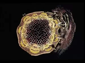
Light Micrograph (LM): A transverse section of a stem of Whisk Fern (Psilotum nudum); Magnification x1200 (on 10)LRDS-182 Light Micrograph (LM): A transverse section of a stem of Whisk Fern Magnification x1200 (on 10.5 cm width print) Psilotum nudum David Spears (Last Refuge)
Light Micrograph (LM): transverse section of Prunus Leaf; Magnification x18 (on 10. 5 cm width print)LRDS-181 Light Micrograph (LM): transverse section of Prunus Leaf Magnification x18 (on 10.5 cm width print) Prunus sp David Spears (Last Refuge)
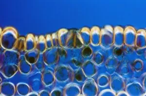
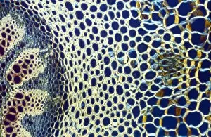
Light Micrograph (LM): transverse section shows Sclerenchyma Ground Tissue in Helianthus stem; Magnification x300LRDS-180 Light Micrograph (LM): transverse section shows Sclerenchyma Ground Tissue in Helianthus stem Magnification x300 (on 10.5 cm width print) Helianthus sp David Spears (Last Refuge)
Light Micrograph (LM): transverse section shows Sclerenchyma Ground Tissue in Helianthus stem; Magnification x1200LRDS-179 Light Micrograph (LM): transverse section shows Sclerenchyma Ground Tissue in Helianthus stem Magnification x1200 (on 10.5 cm width print) Helianthus sp David Spears (Last Refuge)
Light Micrograph (LM): transverse section shows Sclerenchyma Ground Tissue in Helianthus stem; Magnification x1200LRDS-178 Light Micrograph (LM): transverse section shows Sclerenchyma Ground Tissue in Helianthus stem Magnification x1200 (on 10.5 cm width print) Helianthus sp David Spears (Last Refuge)
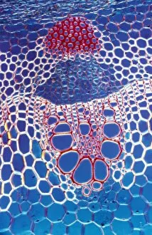
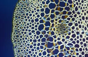
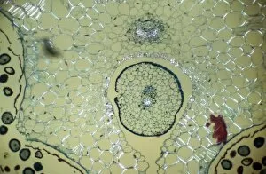
Light Micrograph (LM): A transverse section shows Vascular Bundle in Helianthus stem; Magnification x600 (on 10)LRDS-177 Light Micrograph (LM): A transverse section shows Vascular Bundle in Helianthus stem Magnification x600 (on 10.5 cm width print) Helianthus sp David Spears (Last Refuge)
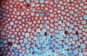
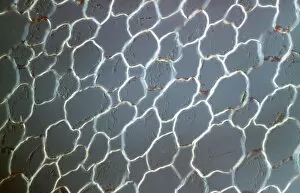
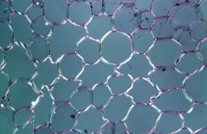
Light Micrograph (LM): transvers section shows Parenchyma Ground Tissue in Buttercup stem; Magnification x600 (on 10)LRDS-176 Light Micrograph (LM): transvers section shows Parenchyma Ground Tissue in Buttercup stem Magnification x600 (on 10.5 cm width print) David Spears (Last Refuge)
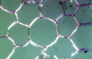
Light Micrograph (LM): transverse section shows Parenchyma or packing tissue from Cucurbita Stem; Magnification x1200LRDS-175 Light Micrograph (LM): transverse section shows Parenchyma or packing tissue from Cucurbita Stem Magnification x1200 (on 10.5 cm width print) David Spears (Last Refuge)
Light Micrograph (LM): tranverse section shows Parenchyma or packing tissue from Cucurbita Stem; Magnification x600LRDS-174 Light Micrograph (LM): tranverse section shows Parenchyma or packing tissue from Cucurbita Stem Magnification x600 (on 10.5 cm width print) David Spears (Last Refuge)
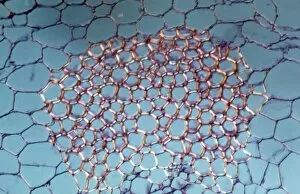
Light Micrograph (LM): A transverse section of a stem of Clubmoss (Lycopodium sp. ); Magnification x600 (on 10)LRDS-173 Light Micrograph (LM): A transverse section of a stem of Clubmoss (Lycopodium sp.) Magnification x600 (on 10.5 cm width print) David Spears (Last Refuge)
Light Micrograph (LM): A transverse section of a stem of Clubmoss (Lycopodium sp. ); Magnification x600 (on 10)LRDS-172 Light Micrograph (LM): A transverse section of a stem of Clubmoss (Lycopodium sp.) Magnification x600 (on 10.5 cm width print) Lycopodium sp
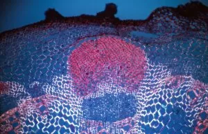
Light Micrograph (LM): A transverse section of a stem of Clover (Trifolium sp. ) with an unidentified parasite attachedLRDS-169 Light Micrograph (LM): A transverse section of a stem of Clover (Trifolium sp.) with an unidentified parasite attached Trifolium sp
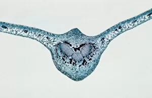
Light Micrograph (LM): transverse section of Dehiscence Lilium Anther shows FilamentLRDS-168 Light Micrograph (LM): transverse section of Dehiscence Lilium Anther shows Filament and Vascular Bundles Magnification x300 (on 10.5 cm width print) David Spears (Last Refuge)