mail_outline sales@mediastorehouse.com
Light Micrograph (LM): A transverse section of a root of Conifer (Pandanus sp. ); Magnification x30 (on 10)LRDS-210 Light Micrograph (LM): A transverse section of a root of Conifer Magnification x30 (on 10.5 cm width print) Pandanus sp
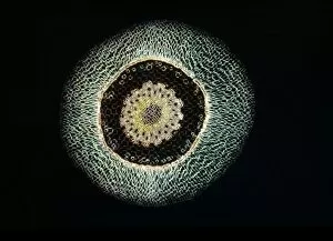
Light Micrograph (LM): A transverse section of an aerial root of Orchid (Dendrobium sp. ); Magnification x30 (on 10)LRDS-209 Light Micrograph (LM): A transverse section of an aerial root of Orchid Magnification x30 (on 10.5 cm width print) Dendrobium sp
Light Micrograph (LM): A transverse section of a leaf of a Tulip (Tulipa sp)LRDS-202 Light Micrograph (LM): A transverse section of a leaf of a Tulip (Tulipa sp.) showing Stomata Magnification x600 (on 10.5 cm width print) Tulipa sp
Light Micrograph (LM): Longitudinal section of old sunflower stem(Helianthus sp)LRDS-199 Light Micrograph (LM): Longitudinal section of old sunflower stem(Helianthus sp.) showing spiral chloroplasts Magnification x1200 (on 10.5 cm width print)
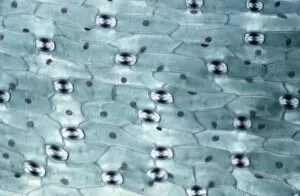
Light Micrograph (LM): A concentration of cells on the epidermis of a plant showing stomataLRDS-197 Light Micrograph (LM): A concentration of cells on the epidermis of a plant showing stomata David Spears (Last Refuge)
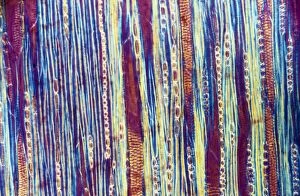
Light Micrograph (LM): Longitudinal section shows Scalariform vessels of Fern; Magnification x1200 (on 10)LRDS-196 Light Micrograph (LM): Longitudinal section shows Scalariform vessels of Fern Magnification x1200 (on 10.5 cm width print) David Spears (Last Refuge)
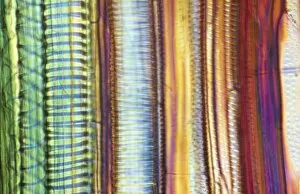
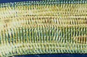
Light Micrograph (LM): A longitudinal section f a Ribes sp. stem; Magnification x600 (on 10. 5 cm width print)LRDS-194 Light Micrograph (LM): A longitudinal section of a Ribes sp. stem Magnification x600 (on 10.5 cm width print) Ribes sp
Light Micrograph (LM): transverse section of Prunus Leaf; Magnification x18 (on 10. 5 cm width print)LRDS-181 Light Micrograph (LM): transverse section of Prunus Leaf Magnification x18 (on 10.5 cm width print) Prunus sp David Spears (Last Refuge)
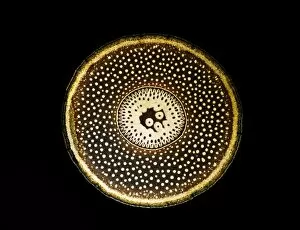
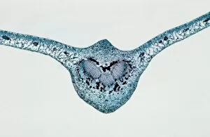
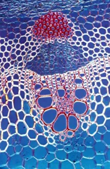
Light Micrograph (LM): A transverse section shows Vascular Bundle in Helianthus stem; Magnification x600 (on 10)LRDS-177 Light Micrograph (LM): A transverse section shows Vascular Bundle in Helianthus stem Magnification x600 (on 10.5 cm width print) Helianthus sp David Spears (Last Refuge)
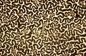
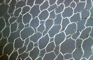
Light Micrograph (LM): transvers section shows Parenchyma Ground Tissue in Buttercup stem; Magnification x600 (on 10)LRDS-176 Light Micrograph (LM): transvers section shows Parenchyma Ground Tissue in Buttercup stem Magnification x600 (on 10.5 cm width print) David Spears (Last Refuge)
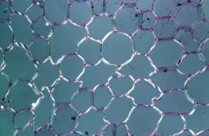
Light Micrograph (LM): tranverse section shows Parenchyma or packing tissue from Cucurbita Stem; Magnification x600LRDS-174 Light Micrograph (LM): tranverse section shows Parenchyma or packing tissue from Cucurbita Stem Magnification x600 (on 10.5 cm width print) David Spears (Last Refuge)
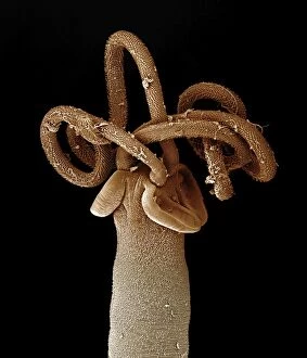
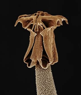
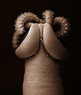
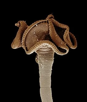
Tapeworm from shark CHI0876LRDS-161 Tapeworm from shark David Spears (Last Refuge) / ardea.com Last Refuge Please note that prints are for personal display purposes only and may not be reproduced in any way
Tapeworm from shark CHI0882LRDS-160 Tapeworm from shark David Spears (Last Refuge) / ardea.com Last Refuge Please note that prints are for personal display purposes only and may not be reproduced in any way
Tapeworm from shark CHI0874LRDS-159 Tapeworm from shark David Spears (Last Refuge) / ardea.com Last Refuge Please note that prints are for personal display purposes only and may not be reproduced in any way
Tapeworm from shark CHI0868LRDS-156 Tapeworm from shark David Spears (Last Refuge) / ardea.com Last Refuge Please note that prints are for personal display purposes only and may not be reproduced in any way
Tapeworm from shark CHI0884LRDS-155 Tapeworm from shark David Spears (Last Refuge) / ardea.com Last Refuge Please note that prints are for personal display purposes only and may not be reproduced in any way
Scanning Electron Micrograph: Woodboring Weevil, Magnification x 222 (A4 size: 29. 7 cm width)LRDS-147 Woodboring Weevil Scanning Electron Micrograph Euophryum confine Magnification x 222 (A4 size: 29.7 cm width) Coloured by hand to enhance natural features
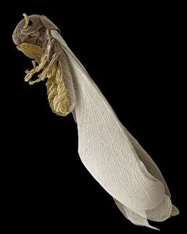
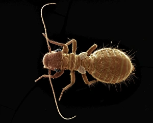
Scanning Electron Micrograph (SEM): Termite, Magnification x 30 (A4 size: 29. 7 cm width)LRDS-145 Termite Scanning Electron Micrograph (SEM) Reticulitermes lucifugus Magnification x 30 (A4 size: 29.7 cm width) Coloured by hand to enhance natural features
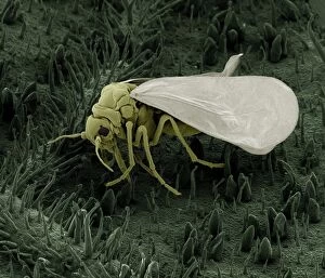
Scanning Electron Micrograph (SEM): Whitefly, Magnification x 130 (A4 size: 29. 7 cm width)LRDS-142 Whitefly, Scanning Electron Micrograph (SEM) Trialeurodes vaporariorum Magnification x 130 (A4 size: 29.7 cm width) Coloured by hand to enhance natural features
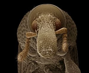
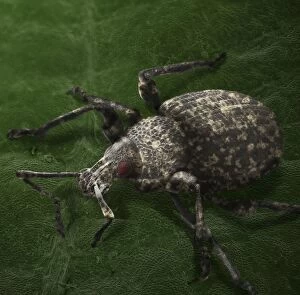
Scanning Electron Micrograph (SEM): Vine Weevil, Magnification x 35 (A4 size: 29. 7 cm width)LRDS-141 Vine Weevil Scanning Electron Micrograph (SEM) Otiorhynchus sulcatus Magnification x 35 (A4 size: 29.7 cm width) Coloured by hand to enhance natural features
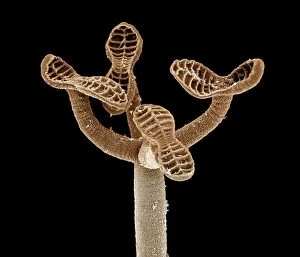
Scanning Electron Micrograph (SEM): Spectacle Moth, Magnification x 200 (A4 size: 29. 7 cm width)LRDS-113 Spectacle Moth - caterpillars hatching Scanning Electron Micrograph (SEM) Abrostola tripartita Magnification x 200 (A4 size: 29.7 cm width) Coloured by hand to enhance natural features
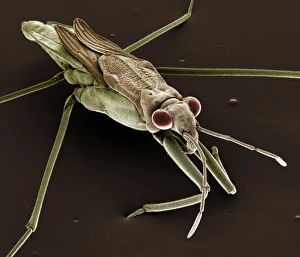
Scanning Electron Micrograph (SEM): Pond Skater, Magnification x 40 (A4 size: 29. 7 cm width)LRDS-111 Pond Skater Scanning Electron Micrograph (SEM) Gerris lacustris Magnification x 40 (A4 size: 29.7 cm width) Coloured by hand to enhance natural features
Scanning Electron Micrograph (SEM): Psocid, Magnification x 100 (A4 size: 29. 7 cm width)LRDS-104 Psocid Scanning Electron Micrograph (SEM) Lepinotus sp. Magnification x 100 (A4 size: 29.7 cm width) Coloured by hand to enhance natural features
Feather Louse - on feather. Magnification x10JLMO-2353 Feather Louse - on feather. Magnification x10 Nirmus merulensis John Mason Please note that prints are for personal display purposes only and may not be reproduced in any way
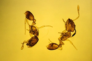
Pharaoh's Ants Magnification x10JLMO-2351 Pharaoh's Ants Magnification x10 Monomorium pharaonis John Mason Please note that prints are for personal display purposes only and may not be reproduced in any way
Anwyl Bromeliads - water is trapped under the scaly on the surface of the plantPPG-1651 Anwyl Bromeliads - water is trapped under the scaly on the surface of the plant Tillandia streptophylla Pascal Goetgheluck Please note that prints are for personal display purposes only
Bladderwort - Bladder-like sac, ready to suck a worm in. South AfricaPPG-1650 Bladderwort - bladder-like sac, ready to suck a worm in South Africa Utricularia sandersonii Pascal Goetgheluck Please note that prints are for personal display purposes only
Drosera pulchella rosea'. Drosera. Greenfly caught by the tentacles. (Electronic Scanning Microscopy)PPG-1644 Drosera pulchella rosea, Greenfly caught by the tentacles Drosera pulchella rosea Electronic Scanning Microscopy Pascal Goetgheluck Please note that prints are for personal display purposes
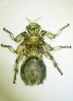
Sheep Ked / TickJC-600 INSECTS - Sheep Ked / Sheep Tick magnified x8 Melophagus ovina John Clegg Please note that prints are for personal display purposes only and may not be reproduced in any way
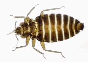
Bed BugJC-598 Bed Bug - male; x 3 magnification Cimex lectularius John Clegg Please note that prints are for personal display purposes only and may not be reproduced in any way